. 2019 Jul 23;19(1):149.
doi: 10.1186/s12862-019-1472-6.

A-to-I editing of Malacoherpesviridae RNAs supports the antiviral role of ADAR1 in mollusks

Affiliations

A-to-I editing of Malacoherpesviridae RNAs supports the antiviral role of ADAR1 in mollusks

Umberto Rosani et al. BMC Evol Biol. .

Abstract

Background: Adenosine deaminase enzymes of the ADAR family are conserved in metazoans. They convert adenine into inosine in dsRNAs and thus alter both structural properties and the coding potential of their substrates. Acting on exogenous dsRNAs, ADAR1 exerts a pro- or anti-viral role in vertebrates and Drosophila.

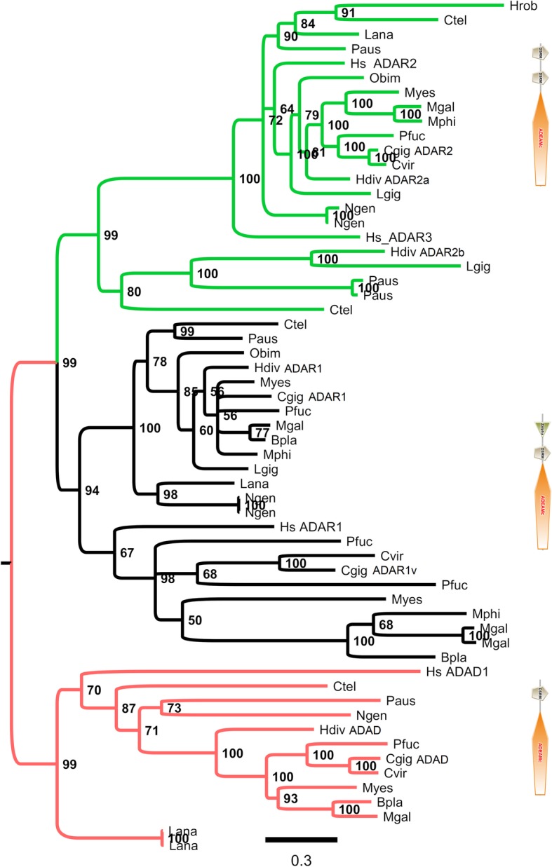
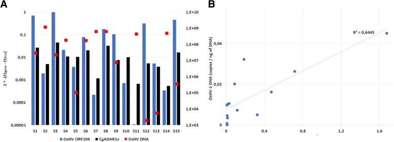
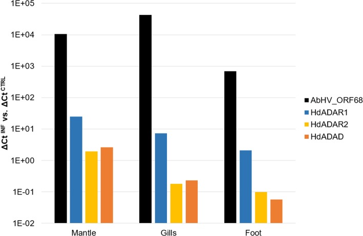
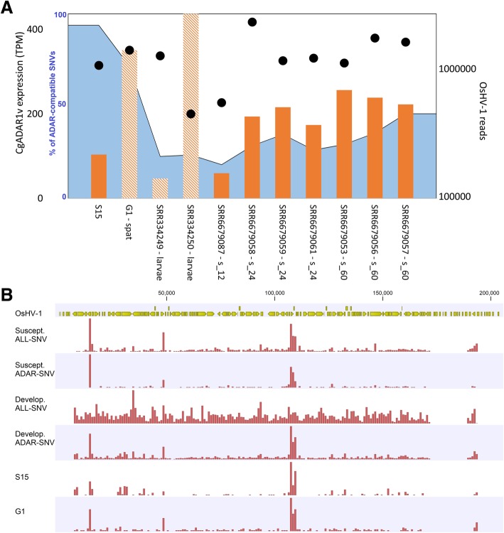
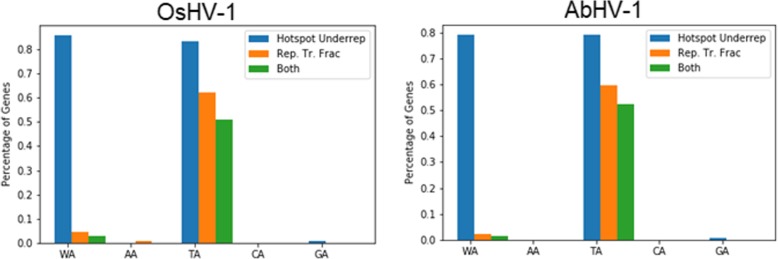
Results: We traced 4 ADAR homologs in 14 lophotrochozoan genomes and we classified them into ADAD, ADAR1 or ADAR2, based on phylogenetic and structural analyses of the enzymatic domain. Using RNA-seq and quantitative real time PCR we demonstrated the upregulation of one ADAR1 homolog in the bivalve Crassostrea gigas and in the gastropod Haliotis diversicolor supertexta during Ostreid herpesvirus-1 or Haliotid herpesvirus-1 infection. Accordingly, we demonstrated an extensive ADAR-mediated editing of viral RNAs. Single nucleotide variation (SNV) profiles obtained by pairing RNA- and DNA-seq data from the viral infected individuals resulted to be mostly compatible with ADAR-mediated A-to-I editing (up to 97%). SNVs occurred at low frequency in genomic hotspots, denoted by the overlapping of viral genes encoded on opposite DNA strands. The SNV sites and their upstream neighbor nucleotide indicated the targeting of selected adenosines. The analysis of viral sequences suggested that, under the pressure of the ADAR editing, the two Malacoherpesviridae genomes have evolved to reduce the number of deamination targets.

Conclusions: We report, for the first time, evidence of an extensive editing of Malacoherpesviridae RNAs attributable to host ADAR1 enzymes. The analysis of base neighbor preferences, structural features and expression profiles of molluscan ADAR1 supports the conservation of the enzyme function among metazoans and further suggested that ADAR1 exerts an antiviral role in mollusks.

Keywords: A-to-I editing; ADAR; AbHV-1; Abalones; Antiviral responses; Malacoherpesvirus; Mollusks; OsHV-1; Oysters; RNA editing.


Conflict of interest statement

The authors declare that they have no competing interests.

Figures

Fig. 1
Fig. 1
Bayesian phylogenesis of ADAR proteins from several lophotrochozoans and Homo sapiens (Hs). The phylogenetic tree is based on the multiple alignment of the catalytic domain provided as Additional file 1. Green, black and red colors denote the ADAR2, ADAR1 and ADAD clusters, respectively, while the typical domain construction of these proteins is reported on the right. Posterior probability values are indicated at each node. Lophotrochozoan species names were abbreviated to 4 letters (e.g. Crassostrea gigas, Cgig)
Fig. 2
Fig. 2
In silico structure model of the CgADAR1 deamination domain. a. Superimposition of the modeled structure of CgADAR1 (green cartoon) to the template HsADAR2 (pale orange cartoon) in complex with dsRNA (PDB ID: 5HP3). The active sites with the RNA flipped base are framed into the black box. b. Magnification of the superimposed active sites. Important residues for the protein’s activity and dsRNA binding, and mutated ones in ADAR1s respect to ADAR2s are represented in sticks. Surface representation of HsADAR2 (c) and CgADAR1 (d) active sites in the dsRNA bound state (represented in dark gray sticks). White arrows indicate the RNA phosphate groups (3′ left, 5′ right) of the flipped, deaminated base, anchored to conserved HsADAR2 residues which are mutated in CgADAR1. Worthy of note is the different steric hindrance and charge distribution of the active sites due to these mutations
Fig. 3
Fig. 3
a. Expression values of OsHV-1 ORF104 (blue) and CgADAR1v (black) in 15 individual oysters (S1-S15) deployed in the lagoon of Goro (North Adriatic Sea, Italy, 2016). qRT-PCR expression data were normalized against CgEl1α. The OsHV-1 DNA copies per ng of total DNA (red points) detected in the same samples are also reported in log10 scale (values on the secondary Y axis). b. Correlation between OsHV-1 ORF104 and CgADAR1v expression values
Fig. 4
Fig. 4
qRT-PCR expression analysis of Haliotis diversicolor supertexta ADARs. For AbHV-1 ORF68, HdADAR1, HdADAR2 and HdADAD the ratio in log10 scale of the deltaCt between viral infected and control samples are reported for the mantle, gill and foot tissues of one abalone (M49)
Fig. 5
Fig. 5
Sequence nucleotide variation (SNV) profiles detected in the viral RNAs obtained from Malacoherpesviridae-infected mollusks. Relative frequency of each possible sequence change based on OsHV-1-PT RNA (one oyster sample, black) and AbHV-1 RNA (three abalone samples collected at 60 hpi; red, yellow and orange)
Fig. 6
Fig. 6
Frequency of different nucleotides (A, C, G, T) at the 5′ and 3′ positions flanking the ADAR-edited base (A) in OsHV-1 and AbHV-1 genomes
Fig. 7
Fig. 7
ADAR editing of OsHV-1 RNA. a. The expression levels of CgADAR1v (TPM, orange bars), the number of OsHV-1 reads (secondary Y-axis in log scale, black circles) and the percentage of ADAR-compatible SNVs over the total number of detected SNVs (blue area depicted on 0–100 scale) are reported for 11 virus-infected oyster samples showing at least 200,000 viral reads. The samples pertaining to pooled individual are depicted by dashed bars. b. Hotspots of ADAR1 editing sites as mapped on the OsHV-1 genome. Viral ORFs and their coding directionality are in yellow. The genome distribution of total and ADAR-compatible SNVs are reported for the susceptible oyster family (12, 24 and 60 hpi samples) and for the developmental stages (two samples). For comparison, the distribution of ADAR-compatible SNVs were also reported for the S15 and G1 samples
Fig. 8
Fig. 8
Under-representation and replacement transition fractions for OsHV-1 and AbHV-1. The blue bars denote the percentage of genes with an under-representation P-value < 0.05 (see Methods); the orange bars denote the percentage of genes with a replacement transition fraction (Rep. Tr. Frac.) P-value > 0.95; the green bars denote the percentage of genes with both the aforementioned P-values. All P-values were corrected using the Benjamini-Hochberg P-value adjustment

References

    1. Koonin EV, Dolja VV. A virocentric perspective on the evolution of life. Curr Opin Virol. 2013;3:546–557. doi: 10.1016/j.coviro.2013.06.008. - DOI - PMC - PubMed
    1. tenOever BR. The Evolution of Antiviral Defense Systems. Cell Host Microbe. 2016;19:142–149. doi: 10.1016/j.chom.2016.01.006. - DOI - PubMed
    1. Aguado LC, Schmid S, May J, Sabin LR, Panis M, Blanco-Melo D, Shim JV, Sachs D, Cherry S, Simon AE, et al. RNase III nucleases from diverse kingdoms serve as antiviral effectors. Nature. 2017;547:114–117. doi: 10.1038/nature22990. - DOI - PMC - PubMed
    1. Suttle CA. Marine viruses — major players in the global ecosystem. Nat Rev Micro. 2007;5:801–812. doi: 10.1038/nrmicro1750. - DOI - PubMed
    1. Andrade KR, Boratto PPVM, Rodrigues FP, Silva LCF, Dornas FP, Pilotto MR, La Scola B, Almeida GMF, Kroon EG, Abrahão JS. Oysters as hot spots for mimivirus isolation. Arch Virol. 2015;160:477–482. doi: 10.1007/s00705-014-2257-2. - DOI - PubMed

Publication types