Review
. 2019 Jul 23;19(1):176.
doi: 10.1186/s12883-019-1392-5.

Primary intraosseous osteolytic meningioma: a case report and review of the literature

Affiliations
Review

Primary intraosseous osteolytic meningioma: a case report and review of the literature

Sae Min Kwon et al. BMC Neurol. .

Abstract

Background: Primary intraosseous meningioma is a subset of extradural meningioma that arises in the bone, and only a few cases have been reported to date.

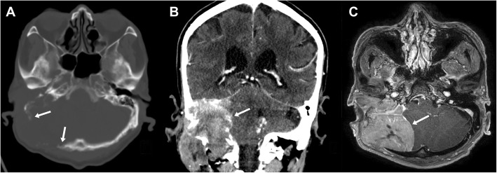
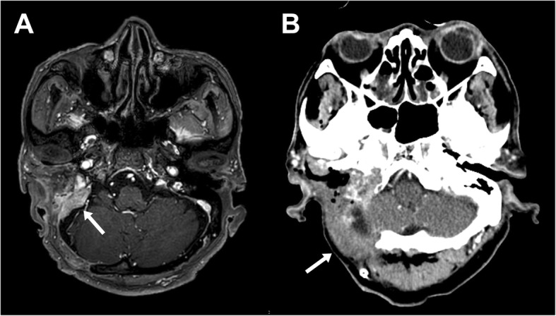
Case presentation: An 80-year-old man presented with decreased hearing on the right side accompanied by a disturbance of balance 10 months prior to admission. Magnetic resonance imaging revealed an 8 × 7 cm osteolytic mass in the right posterior fossa related to the petrous bone, with extension to the cervical region. During surgery, the tumor was found to be located extradurally, with no invasion of the dura. The tumor was removed entirely, apart from a small portion around the jugular foramen to avoid lower cranial nerve injury.

Conclusion: The final diagnosis was primary intraosseous osteolytic meningioma with atypical pathology. Here, we report a rare case of an osteolytic skull lesion in the skull base not invading the dura and with extensive bone destruction.

Keywords: Intraosseous; Meningioma; Osteolysis.


Conflict of interest statement

The authors declare that they have no competing interests.

Figures

Fig. 1
Fig. 1
Preoperative imaging. Axial computed tomography (CT) scan with bone window (a) shows a destructive osteolytic mass lesion in the right temporal-posterior fossa region. Coronal CT scan (b) and magnetic resonance imaging (c) revealed an 8 × 7 cm homogenous mass extending to the cervical region
Fig. 2
Fig. 2
Postoperative magnetic resonance imaging (a) shows a small residual tumor around the jugular foramen, and computed tomography scan (b) demonstrates the sternocleidomastoid muscle which fills the tumor removal space
Fig. 3
Fig. 3
Histopathologic findings of atypical meningioma. The fragmented specimen (a) is seen as grayish-white solid masses. On microscopic examination, the tumor infiltrated the adjacent soft tissue (b, hematoxylin and eosin [H&E] stain, × 20, scale bar = 200 μm) and showed a whorled appearance and multifocal necrosis (c, H&E stain, × 100, scale bar = 100 μm). The tumor cells are composed of spindle cells with prominent nucleoli and ill-defined cytoplasm. Many mitoses are visible (d, H&E stain, × 400, scale bar = 20 μm)

Similar articles

Cited by

References

    1. Agrawal V, Ludwig N, Agrawal A, Bulsara KR. Intraosseous Intracranial Meningioma. Am J Neuroradiol. 2007;28:314–315. - PMC - PubMed
    1. Muzumdar DP, Vengsarkar US, Bhatjiwale MG, Goel A. Diffuse calvarial meningioma: a case report. J Postgrad Med. 2001;47:116. - PubMed
    1. Lang FF, Macdonald OK, Fuller GN, DeMonte F. Primary extradural meningiomas: a report on nine cases and review of the literature from the era of computerized tomography scanning. J Neurosurg. 2000;93:940–950. doi: 10.3171/jns.2000.93.6.0940. - DOI - PubMed
    1. Nicolay S, De Foer B, Bernaerts A, Van Dinther J, Parizel PM. A case of a temporal bone meningioma presenting as a serous otitis media. Acta Radiol Short Rep. 2014;3:2047981614555048. - PMC - PubMed
    1. Hoye SJ, Hoar CS, Murray JE. Extracranial meningioma presenting as a tumor of the neck. Am J Surg. 1960;100:486–489. doi: 10.1016/0002-9610(60)90394-9. - DOI - PubMed

LinkOut - more resources