. 2019 Jul 23;38(1):326.
doi: 10.1186/s13046-019-1325-6.

Anti-PSMA 124I-scFvD2B as a new immuno-PET tool for prostate cancer: preclinical proof of principle

Affiliations

Anti-PSMA 124I-scFvD2B as a new immuno-PET tool for prostate cancer: preclinical proof of principle

B Frigerio et al. J Exp Clin Cancer Res. .

Abstract

Background: Prostate cancer (PCa) is the second leading cause of cancer-related death in the Western population. The use in oncology of positron emission tomography/computed tomography (PET/CT) with emerging radiopharmaceuticals promises accurate staging of primary disease, restaging of recurrent disease and detection of metastatic lesions. Prostate-specific membrane antigen (PSMA) expression, directly related to androgen-independence, metastasis and progression, renders this tumour associate antigen a good target for the development of new radiopharmaceuticals for PET. Aim of this study was to demonstrate in a preclinical in vivo model (PSMA-positive versus PSMA-negative tumours) the targeting specificity and sensitivity of the anti-PSMA single-chain variable fragment (scFv) labelled with 124I.

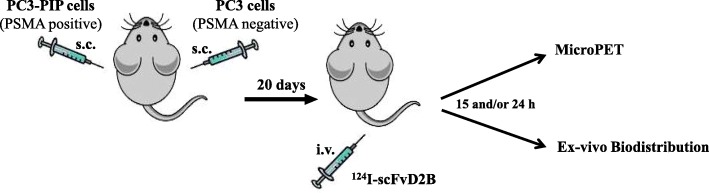
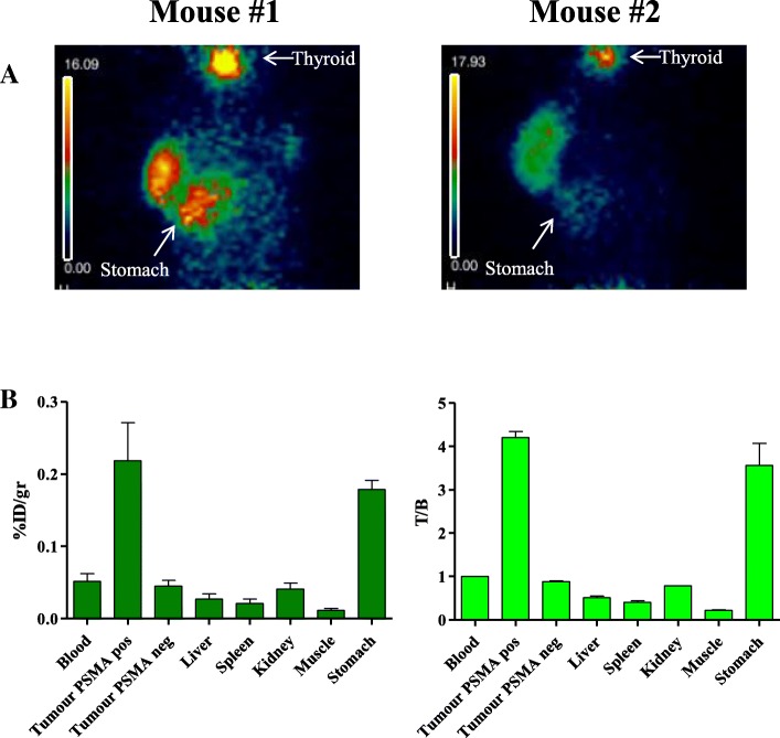
Methods: The 124I-labeling conditions of the antibody fragment scFvD2B were optimized and assessed for purity and immunoreactivity. The specificity of 124I-scFvD2B was tested in mice bearing PSMA-positive and PSMA-negative tumours to assess both ex-vivo biodistribution and immune-PET.

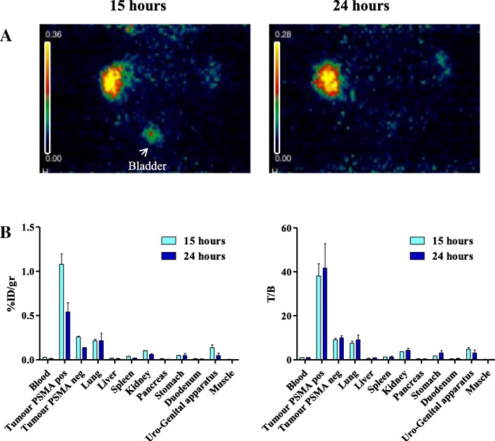
Results: The uptake fraction of 124I-scFvD2B was very high on PSMA positive cells (range 75-91%) and highly specific and immuno-PET at the optimal time point, defined between 15 h and 24 h, provides a specific localization of lesions bearing the target antigen of interest (PSMA positive vs PSMA negative tumors %ID/g: p = 0.0198 and p = 0.0176 respectively) yielding a median target/background ratio around 30-40.

Conclusions: Preclinical in vivo results of our immuno-PET reagent are highly promising. The target to background ratio is improved notably using PET compared to SPECT previously performed. These data suggest that, upon clinical confirmation of sensitivity and specificity, our anti-PSMA 124I-scFvD2B may be superior to other diagnostic modalities for PCa. The possibility to combine in patients our 124I-scFvD2B in multi-modal systems, such as PET/CT, PET/MR and PET/SPECT/CT, will provide quantitative 3D tomographic images improving the knowledge of cancer biology and treatment.

Keywords: 124I; Antibody fragment; PCa; PET; scFv.


Conflict of interest statement

The authors declare no conflicts of interest. The funding agencies had no role in study design and collection, analysis and interpretation of data, nor in writing this paper or the decision to submit it for publication.

Figures

Fig. 1
Fig. 1
Cartoon illustrating the in vivo procedure: 5 × 106 of either PC3-PIP (PSMA positive) or PC3 (PSMA negative) cells were injected subcutaneously (s.c.) in the flank of mice. 20 days later, 124I-scFvD2B was administered i.v. and after 15–24 h both ImmunoPET imaging and ex vivo biodistribution were performed
Fig. 2
Fig. 2
ImmunoPET imaging and ex vivo biodistribution analysis with 124I-scFvD2B of experiment #1: a Immuno-PET images after intravenous administration of 3.7 MBq 124IscFvD2B (166.5 MBq/mg) in two mice. PC3 (PSMA negative, right flank) and PC3-PIP (PSMA positive, left flank) tumours evaluated 24 h post injection. Stomach and Thyroid are indicated by an arrow. The imaging is so clean in the irrelevant organ that it is barely visible the shape of the mice. b Ex vivo biodistribution analysis in PC3 and PC3-PIP tumours in the same two mice. Uptake and retention were measured in different organs and illustrated both as decay-adjusted percentage of the injected dose per gram of tissue (% ID/g) and T/B. The background activity observed in the stomach was probably due to the free 124I or the possible ingestion of the sawdust contaminated by the radioactive fraction present in the mice urine since was not present in the fourth experiment done in a larger number of animals (Fig. 3b)
Fig. 3
Fig. 3
ImmunoPET imaging and ex vivo biodistribution analysis with 124I-scFvD2B of experiment #4. a One representative Immuno-PET images after intravenous administration of 7.4 MBq 124IscFvD2B (433 MBq/mg). PC3 (PSMA negative, right flank) and PC3-PIP (PSMA positive, left flank) tumours evaluated 15 and 24 h post injection. Bladder is also indicated by an arrow. The imaging is so clean in the irrelevant organ that it is barely visible the shape of the mice. b Ex vivo biodistribution analysis in PC3 and PC3-PIP tumour bearing mice evaluated 15 h (2 mice) and 24 h (3 mice) post injection of 124I-scFvD2B. Uptake and retention were measured in different organs and illustrated both as decay-adjusted percentage of the injected dose per gram of tissue (% ID/g) and T/B

Similar articles

Cited by

References

    1. Zhou CK, Check DP, Lortet-Tieulent J, Laversanne M, Jemal A, Ferlay J, et al. Prostate cancer incidence in 43 populations worldwide: an analysis of time trends overall and by age group. Int J Cancer. 2016;138:1388–1400. doi: 10.1002/ijc.29894. - DOI - PMC - PubMed
    1. Mottet N, Bellmunt J, Bolla M, Briers E, Cumberbatch MG, De Santis M, et al. EAU-ESTRO-SIOG guidelines on prostate Cancer. Part 1: screening, diagnosis, and local treatment with curative intent. Eur Urol. 2017;71:618–629. doi: 10.1016/j.eururo.2016.08.003. - DOI - PubMed
    1. Xu KM, Chen RC, Schuster DM, Jani AB. Role of novel imaging in the management of prostate cancer. Urol Oncol: Seminars and Original Investigations. 2019. - PubMed
    1. Yang H, Liu T, Wang X, Xu Y, Deng S. Diagnosis of bone metastases: a meta-analysis comparing 18FDG PET, CT, MRI and bone scintigraphy. Eur Radiol. 2011;21:2604–2617. doi: 10.1007/s00330-011-2221-4. - DOI - PubMed
    1. de Rooij M, Hamoen EHJ, Witjes JA, Barentsz JO, Rovers MM. Accuracy of magnetic resonance imaging for local staging of prostate Cancer: a diagnostic meta-analysis. Eur Urol. 2016;70:233–245. doi: 10.1016/j.eururo.2015.07.029. - DOI - PubMed

MeSH terms