. 2019 Oct;9(10):1438-1451.
doi: 10.1158/2159-8290.CD-19-0393. Epub 2019 Jul 23.

A Mutation in Histone H2B Represents a New Class of Oncogenic Driver

Affiliations

A Mutation in Histone H2B Represents a New Class of Oncogenic Driver

Richard L Bennett et al. Cancer Discov. 2019 Oct.

Abstract

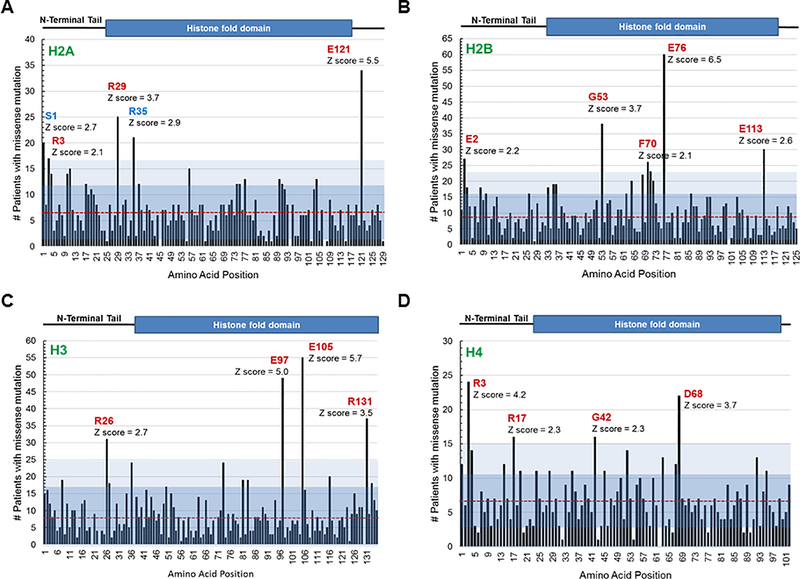
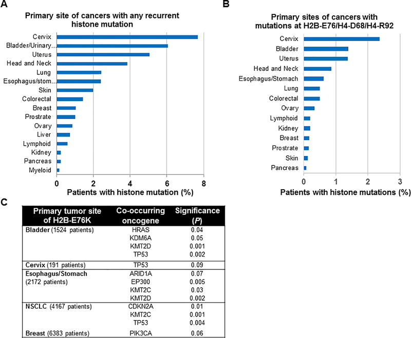
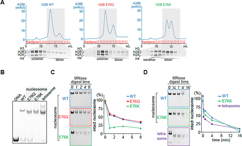
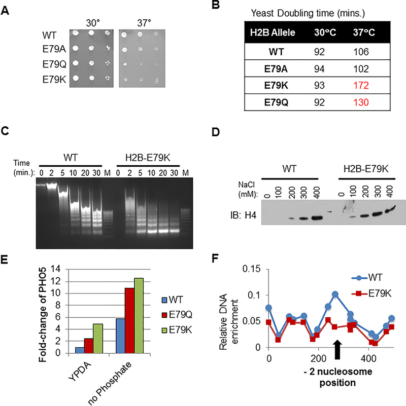
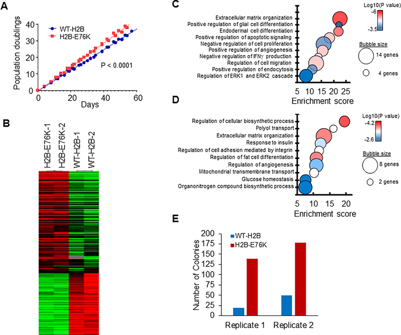
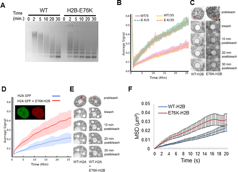
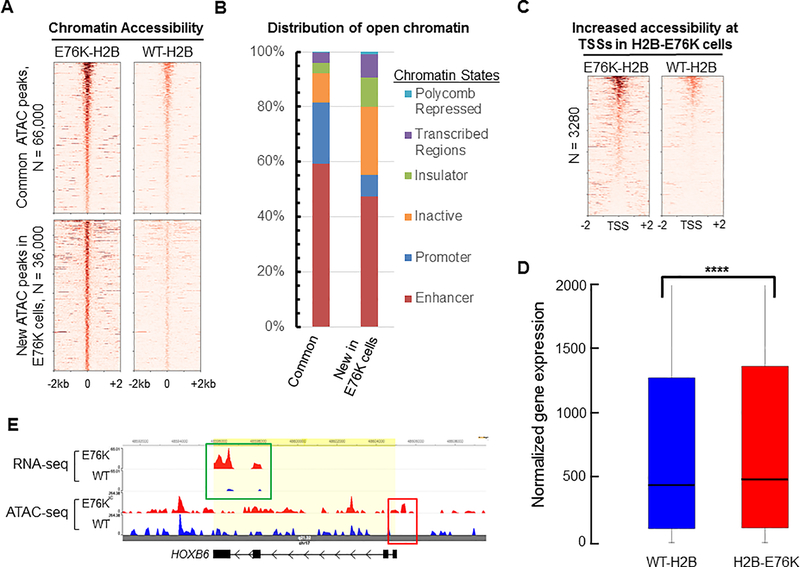
By examination of the cancer genomics database, we identified a new set of mutations in core histones that frequently recur in cancer patient samples and are predicted to disrupt nucleosome stability. In support of this idea, we characterized a glutamate to lysine mutation of histone H2B at amino acid 76 (H2B-E76K), found particularly in bladder and head and neck cancers, that disrupts the interaction between H2B and H4. Although H2B-E76K forms dimers with H2A, it does not form stable histone octamers with H3 and H4 in vitro, and when reconstituted with DNA forms unstable nucleosomes with increased sensitivity to nuclease. Expression of the equivalent H2B mutant in yeast restricted growth at high temperature and led to defective nucleosome-mediated gene repression. Significantly, H2B-E76K expression in the normal mammary epithelial cell line MCF10A increased cellular proliferation, cooperated with mutant PIK3CA to promote colony formation, and caused a significant drift in gene expression and fundamental changes in chromatin accessibility, particularly at gene regulatory elements. Taken together, these data demonstrate that mutations in the globular domains of core histones may give rise to an oncogenic program due to nucleosome dysfunction and deregulation of gene expression. SIGNIFICANCE: Mutations in the core histones frequently occur in cancer and represent a new mechanism of epigenetic dysfunction that involves destabilization of the nucleosome, deregulation of chromatin accessibility, and alteration of gene expression to drive cellular transformation.See related commentary by Sarthy and Henikoff, p. 1346.This article is highlighted in the In This Issue feature, p. 1325.


Conflict of interest statement

Disclosures: The authors have no conflicts of interest to declare

Figures

Figure 1.
Figure 1.. Survey of the most frequently found canonical histone mutations in cancer patient samples.
A cross cancer mutation summary was performed using the cBioPortal to search a total of 41,738 non-redundant patient samples across all cancer types. The number of patients reported to have a missense mutation at each amino acid residue position across histone paralogs was graphed. A red line denotes the average number of mutations for each histone across all paralogs, and blue shaded regions indicate the first two standard deviations from the average. Location of the cumulative missense mutations found in: (A) 15 canonical H2A genes (HIST1H2AA/B/C/D/E/G/H/I/J/K/L/M, HIST2H2AB/C, HIST3H2A), (B) 18 canonical H2B genes (HIST1H2BA/B/C/D/E/F/G/H/I/J/K/L/M/N/O, HIST2H2BE/F, HIST3H2BB), (C) 12 canonical H3 genes (HIST1H3A/B/C/D/E/F/G/H/I/J, HIST2H3D, HIST3H3), and (D) 14 canonical H4 genes (HIST1H4A/B/C/D/E/F/G/H/I/J/K/L, HIST2H4A, HIST4H4) across all cancer patient samples. The Z scores indicate the most frequently found mutations (Z > 2) in each histone. Amino acids displayed in red were significantly more frequent in cBioPortal than the general SNP database.
Figure 2.
Figure 2.. Frequency of recurrent histone missense mutations in cancer.
The cBioPortal was queried to determine the frequency of histone mutations in cancer patient samples at each primary site. (A) The frequency for any of the eighteen most significantly recurring histone missense mutations (Z score > 2) in each primary cancer site. (B) The frequency of missense mutations at either H2B glutamate 76 (H2B-E76), H4 aspartate (H4-D68) or H4 arginine (H4-R92), which destabilizes the histone octamer at a specific structural region, in cancers at each primary site. (C) The co-occurrence of the H2B-E76K mutation with mutations in the most common oncogenes found in each cancer type (for cancers with 3 or more patients with H2B-E76K) was assessed by a randomization test through 10,000 rounds of randomization.
Figure 3:
Figure 3:. The E76K mutation in H2B destabilizes the histone octamer and fails to protect the nucleosome from nuclease treatment in vitro.
(A) The H2B-E76K mutant was unable to form stable octamers in vitro. Recombinant human histones (H2A, H2B, H3, and H4) were mixed and histone octamers resolved from (H3/H4)2 tetramers, H2A/H2B dimers and free histones by gel filtration chromatography. (B) Nucleosomes were reconstituted by mixing equimolar amounts of DNA (147bp) and octamers or in the case of E76K of tetramers and dimers (1:2 molar ratio) and resolved by Native PAGE. Nucleosomes containing H2B-E76K and E76Q have an altered migration pattern, intermediate between a tetrasome and a WT nucleosome. (C) Micrococcal nuclease (MNase) sensitivity assay performed on nucleosomes made with WT, E76Q and E76K H2B mutants shows more rapid digestion of E76K containing nucleosomes than those with WT H2B. A time course by gel (left) and densitometry quantification (right) of intact nucleosomes following MNase treatment. (D) The MNase susceptibility of E76K nucleosomes is distinct from nucleosomes formed only with tetrasomes.
Figure 4.
Figure 4.. Expression of mutant H2B in yeast destabilizes nucleosomes, deregulates gene expression and reduces nucleosome occupancy at the PHO5 promoter.
WT or E79K H2B (analogous to human H2B-E76K) was expressed in S. Cerevisiae. (A) Yeast cells expressing H2B-E79K are temperature sensitive. Limiting dilutions of yeast expressing WT, E79A, E79Q or E79K were plated and incubated at 30°C or 37°C. Cell growth was evaluated after 1 day. (B) Yeast doubling time is significantly increased in cells expressing E79K-H2B at 37°C. (C) Time course of MNase sensitivity from spheroplasted yeast grown in rich media. M, marker. (D) Chromatin pellets were extracted with increasing concentrations of salt as indicated. Immuno-blotting of the soluble fraction was performed with antibody to H4. (E) Cells expressing WT, E79Q or E79K H2B were maintained in either rich media (YPDA) or phosphate-free media and expression of the phosphate-inducible PHO5 gene was measured by RT-PCR. (F) Nucleosome scanning assay of the PHO5 promoter from cells expressing either WT or E79K grown in rich media. Chromatin was digested with MNase, mononucleosomal DNA was purified and MNase protection was determined by qPCR. H2B occupancy at −2 nucleosome position of PHO5 is reduced in E79K cells as indicated by the arrow.
Figure 5.
Figure 5.. Expression of H2B-E76K alters growth properties and gene expression in human mammary epithelial cells.
(A) Proliferation of MCF10A transduced with lentivirus expressing either WT or H2B-E76K was measured every 3 to 4 days. The results from three different infections are shown. Linear regression analysis was used to calculate statistical significance. (B) Heat maps depicting differential gene expression between MCF10A cells expressing either WT or H2B-E76K. (C and D) Bubble plots of gene ontology analysis identify biological processes significantly different in cells expressing H2B-E76K compared to cells expressing WT H2B. Bubble area is relative to the number of genes identified in each classification. Ontology of genes with increased (C) or decreased (D) expression in cells expressing H2B-E76K. (E) MCF10A cells expressing WT H2B or H2B-E76K and PIK3CA(H1047R) were grown in soft agar for 14 days and colonies counted.
Figure 6.
Figure 6.. H2B-E76K fundamentally alters chromatin structure and dynamics.
(A) Micrococcal nuclease (MNase) assay with nuclei of MCF10A cells stably expressing either WT or H2B-E76K demonstrate that E76K expression significantly increases sensitivity to MNase. Digest efficiency was visualized by agarose gel. (B) Fluorescence recovery after photobleaching (FRAP) analysis demonstrates that H2B-E76K has significantly faster chromatin dynamics than WT. FRAP analysis was carried out after induction of either WT or E76K mutant H2B-GFP fusions for 5 or 35 days in MCF10A cells. Dashed lines represent cells expressing H2B-E76K, solid lines represent data from cells expressing inducible GFP-tagged WT H2B (n = 10 cells). (C) Representative FRAP assay pre-bleach, bleach and post-bleach images of nuclei expressing GFP-tagged WT or H2B-E76K indicate faster fluorescent recovery in cells expressing E76K. (D) FRAP analysis of histone H2A-GFP dynamics in MCF10A cells expressing only H2A-GFP (n = 20 cells) or both H2A-GFP and a mutant H2B E76K mCherry fusion (n = 25 cells) with standard deviation envelopes. Inset demonstrates co-expression of H2A GFP and E76K mCherry. (E) Representative pre-bleach, bleach and post-bleach images of H2A GFP in WT MCF10A cells or in cells co-expressing mCherry tagged H2B-E76K. (F) 100nm particles injected into the nucleus had significantly increased mean square displacement (MSD) over time in MCF10A cells stably expressing exogenous H2B-E76K (red) compared to cells expressing WT H2B (blue). N=68 (WT) and N=67 (E76K) cells were analyzed. Error bars represent SEM.
Figure 7.
Figure 7.. Expression of H2B E76K alters chromatin accessibility in MCF10A cells.
(A) Heat maps of ATAC-Seq peaks from MCF10A cells expressing either WT H2B or H2B-E76K found within a ± 2kb window, ordered by highest ATAC-Seq signal to lowest. Peaks found in both cell lines are listed as common peaks. Peaks significantly enriched in cells expressing H2B-E76K are denoted as new peaks in E76K. (B) The distribution of ATAC-Seq peaks among six broad classes of chromatin states was performed using the encode HMEC ChromHMM data set. (C) Heat maps of ATAC-Seq reads within +/− 2 kb of transcription start sites (TSS) for genes with significantly increased chromatin accessibility at promoter regions of MCF10A cells expressing H2B-E76K compared to cells expressing WT-H2B. (D) Box and whiskers plot for gene expression of the 3280 genes that have increased ATAC-Seq signal at the TSS in cells expressing H2B-E76K compared to expression of those genes in cells expressing WT-H2B. *** indicates that P < 0.001. (E) Genome browser tracks of a representative gene (HOXB6) with increased chromatin accessibility at the TSS in cells expressing H2B-E76K (red box) that corresponds with increased expression (green box).

Comment in

  • Bringing Oncohistones into the Fold.
    Sarthy JF, Henikoff S. Sarthy JF, et al. Cancer Discov. 2019 Oct;9(10):1346-1348. doi: 10.1158/2159-8290.CD-19-0839. Cancer Discov. 2019. PMID: 31575564

References

    1. Lawrence MS, Stojanov P, Mermel CH, Robinson JT, Garraway LA, Golub TR, et al. Discovery and saturation analysis of cancer genes across 21 tumour types. Nature 2014;505(7484):495–501 doi 10.1038/nature12912. - DOI - PMC - PubMed
    1. Kornberg RD, Thomas JO. Chromatin structure; oligomers of the histones. Science 1974;184(4139):865–8. - PubMed
    1. Kornberg RD. Chromatin structure: a repeating unit of histones and DNA. Science 1974;184(4139):868–71. - PubMed
    1. Lorch Y, LaPointe JW, Kornberg RD. Nucleosomes inhibit the initiation of transcription but allow chain elongation with the displacement of histones. Cell 1987;49(2):203–10. - PubMed
    1. Struhl K, Segal E. Determinants of nucleosome positioning. Nature structural & molecular biology 2013;20(3):267–73 doi 10.1038/nsmb.2506. - DOI - PMC - PubMed

Publication types