

Manganese-enhanced MRI of the myocardium
- PMID: 31337670
- PMCID: PMC6855794
- DOI: 10.1136/heartjnl-2019-315227
Manganese-enhanced MRI of the myocardium
Abstract
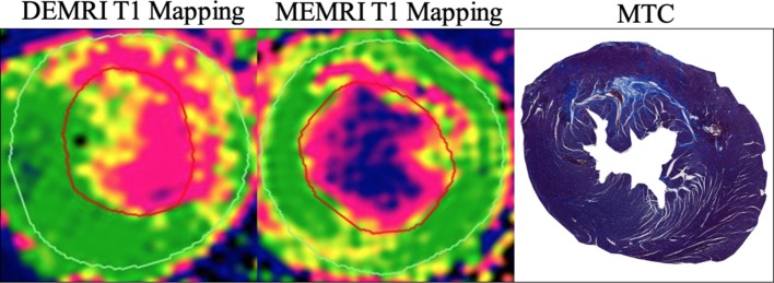
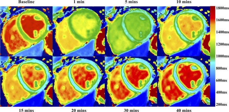
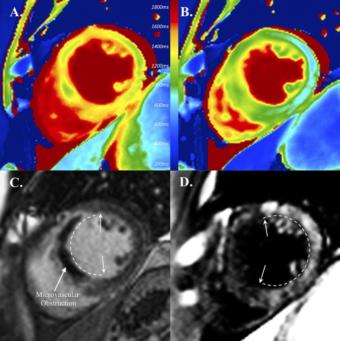
Gadolinium-based contrast media are widely used in cardiovascular MRI to identify and to highlight the intravascular and extracellular space. After gadolinium, manganese has the second highest paramagnetic moment and was one of the first MRI contrast agents assessed in humans. Over the last 50 years, manganese-enhanced MRI (MEMRI) has emerged as a complementary approach enabling intracellular myocardial contrast imaging that can identify functional myocardium through its ability to act as a calcium analogue. Early progress was limited by its potential to cause myocardial depression. To overcome this problem, two clinical formulations of manganese were developed using either chelation (manganese dipyridoxyl diphosphate) or coadministration with a calcium compound (EVP1001-1, Eagle Vision Pharmaceuticals). Preclinical studies have demonstrated the efficacy of MEMRI in quantifying myocardial infarction and detecting myocardial viability as well as tracking altered contractility and calcium handling in cardiomyopathy. Recent clinical data suggest that MEMRI has exciting potential in the quantification of myocardial viability in ischaemic cardiomyopathy, the early detection of abnormalities in myocardial calcium handling, and ultimately, in the development of novel therapies for myocardial infarction or heart failure by actively quantifying viable myocardium. The stage is now set for wider clinical translational study of this novel and promising non-invasive imaging modality.
Keywords: MEMRI; manganese-enhanced MRI; viability.
© Author(s) (or their employer(s)) 2019. Re-use permitted under CC BY. Published by BMJ.
Conflict of interest statement
Competing interests: None declared.
Figures
References
-
- Kanda T, Fukusato T, Matsuda M, et al. . Gadolinium-based contrast agent accumulates in the brain even in subjects without severe renal dysfunction: evaluation of autopsy brain specimens with inductively coupled plasma mass spectroscopy. Radiology 2015;276:228–32. 10.1148/radiol.2015142690 - DOI - PubMed
-
- Kang YS, Gore JC. Studies of tissue NMR relaxation enhancement by manganese. Dose and time dependences. Invest Radiol 1984;19:399–407. - PubMed
-
- Santamaria AB. Manganese exposure, essentiality & toxicity. Indian J Med Res 2008;128:484–500. - PubMed
-
- Lauterbur PC. Image formation by induced local interactions. Examples employing nuclear magnetic resonance. 1973. Clin Orthop Relat Res 1989;242:190–1. - PubMed
Publication types
MeSH terms
Substances
Grants and funding
LinkOut - more resources
Full Text Sources
Medical