. 2019 Aug 6;116(32):15895-15900.
doi: 10.1073/pnas.1818547116. Epub 2019 Jul 23.

Perturbation of the interactions of calmodulin with GRK5 using a natural product chemical probe

Affiliations

Perturbation of the interactions of calmodulin with GRK5 using a natural product chemical probe

Tyler S Beyett et al. Proc Natl Acad Sci U S A. .

Abstract

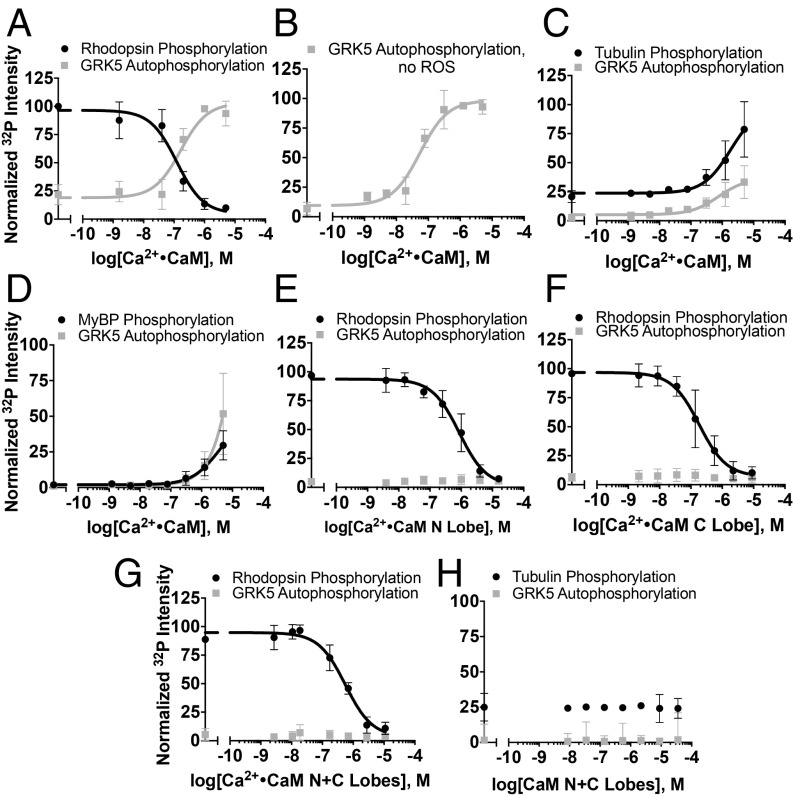
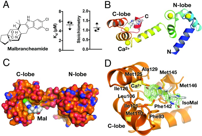
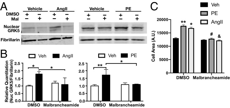
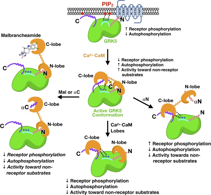
G protein-coupled receptor (GPCR) kinases (GRKs) are responsible for initiating desensitization of activated GPCRs. GRK5 is potently inhibited by the calcium-sensing protein calmodulin (CaM), which leads to nuclear translocation of GRK5 and promotion of cardiac hypertrophy. Herein, we report the architecture of the Ca2+·CaM-GRK5 complex determined by small-angle X-ray scattering and negative-stain electron microscopy. Ca2+·CaM binds primarily to the small lobe of the kinase domain of GRK5 near elements critical for receptor interaction and membrane association, thereby inhibiting receptor phosphorylation while activating the kinase for phosphorylation of soluble substrates. To define the role of each lobe of Ca2+·CaM, we utilized the natural product malbrancheamide as a chemical probe to show that the C-terminal lobe of Ca2+·CaM regulates membrane binding while the N-terminal lobe regulates receptor phosphorylation and kinase domain activation. In cells, malbrancheamide attenuated GRK5 nuclear translocation and effectively blocked the hypertrophic response, demonstrating the utility of this natural product and its derivatives in probing Ca2+·CaM-dependent hypertrophy.

Keywords: G protein-coupled receptor kinase 5; calmodulin; hypertrophy; malbrancheamide.


Conflict of interest statement

The authors declare no conflict of interest.

Figures

Fig. 1.
Fig. 1.
Biophysical characterization of the Ca2+·CaM–GRK5 complex. (A) Representative chromatograms of GRK5 ± Ca2+·CaM (3-fold molar excess) passed over an S200 size exclusion chromatography column. A shift toward a lower elution volume was observed in the presence of Ca2+·CaM. MALS determined the molecular weight of the complex to be 90 kDa (solid line inside the peak), the approximate weight of a 1:1 complex (n = 2). The molecular weights of His-tagged GRK5 and CaM are 70.7 and 19.6 kDa, respectively. (B) SDS/PAGE analysis of the chromatogram peak corresponding to the Ca2+·CaM–GRK5 complex. Both proteins coelute, consistent with complex formation. (C) Reconstructed density at 3 σ calculated via DENSS from SEC–SAXS scattering curves of the complex at 10 mg mL−1. GRK5 (PDB entry 4WNK) with terminal regions from GRK6 (PDB entry 3NYN) and Ca2+·CaM (orange cartoon, PDB entry 5J03) autodocked into the envelope. The regulator of G protein-signaling homology (RH) domain (colored beige), kinase domain (KD; colored green), active-site tether (AST; colored purple), basic phospholipid-binding patch (side-chains shown as spheres), and terminal helices (αN and αC; mauve and purple, respectively) are labeled. The model fits to the RAW SAXS data well (χ2 = 1.07) as assessed by FoXS. (D) Comparison of selected negative-stain class averages of GRK5 alone and the Ca2+·CaM–GRK5 complex. Arrows indicate density attributed to Ca2+·CaM.
Fig. 2.
Fig. 2.
Modulation of substrate phosphorylation by Ca2+·CaM. (A) Rhodopsin in rod outer segments (ROS, 5 µM, light-activated) phosphorylation (IC50 = 120 ± 40 nM) and concurrent GRK5 (50 nM) autophosphorylation (EC50 = 170 ± 70 nM) in the presence of Ca2+·CaM. (B) Stimulation of GRK5 (50 nM) autophosphorylation by Ca2+·CaM (EC50 = 50 ± 20 nM) in the absence of ROS or other substrates. (C) Tubulin (10 µM) phosphorylation in the presence of Ca2+·CaM. Decreased potency of concurrent autophosphorylation is likely due to competition between the C terminus of GRK5 and tubulin. (D) Phosphorylation of myelin basic protein (MyBP, 7 µM) presence of Ca2+·CaM and concurrent autophosphorylation. Inhibition of rhodopsin phosphorylation by the (E) Ca2+·CaM N lobe (IC50 = 860 ± 230 nM), (F) C lobe (IC50 = 180 ± 60 nM), or (G) a 1:1 molar combination of N and C lobes (IC50 = 600 ± 170 nM). (H) Phosphorylation of tubulin is unaffected by the addition of both lobes. All radiometric kinase assays were performed three times and reported as mean ± SD and fit to a sigmoidal dose–response model in GraphPad Prism with the Hill slope constrained to 1.
Fig. 3.
Fig. 3.
Malbrancheamide binds to the C lobe of Ca2+·CaM. (A) Chemical structure of malbrancheamide and ITC-binding results (average ± SD, n = 3). (B) Rainbow ribbon representation of the Ca2+·CaM·malbrancheamide structure in a trans conformation. Ca2+ are shown as yellow spheres (PDB entry 6EEB). (C) Surface representation of Ca2+·CaM (orange corresponding to carbon, blue nitrogen, and red oxygen) with malbrancheamide shown as spheres with white carbon atoms, green chlorine. (D) C-lobe hydrophobic pocket with isomalbrancheamide D (IsoMal) bound (bromine colored brown). Green cage represents simulated annealing |Fo| − |Fc| omit density contoured at 2.5 σ. Side-chains making van der Waals contacts with IsoMal are labeled and are similar to those contacting malbrancheamide (SI Appendix, Fig. S7). Color scheme is the same as in C.
Fig. 4.
Fig. 4.
Effects of malbrancheamide and GRK5 peptides on Ca2+·CaM modulation of GRK5 activity. (A) Rhodopsin (5 µM, light-activated) phosphorylation in the presence of 500 nM Ca2+·CaM and increasing concentrations of malbrancheamide. In these assays, Ca2+·CaM was preincubated with malbrancheamide before the addition of GRK5. (B) Tubulin (5 µM) phosphorylation in the presence of 500 nM Ca2+·CaM and varying concentrations of malbrancheamide. (C) Rhodopsin phosphorylation in the presence of 500 nM Ca2+·CaM and varying concentrations of GRK5 αN (residues 2 to 31, IC50,autophosphorylation = 540 ± 240 nM, EC50,rhodopsin = 5.6 ± 1.6 µM) or (D) αC (residues 546 to 565, IC50 = 1.1 ± 0.7 µM) peptides. (E) Tubulin phosphorylation in the presence of 500 nM Ca2+·CaM and varying concentrations of GRK5 αN (IC50 = 2.4 ± 1.7 µM) or (F) αC peptides (IC50 = 500 ± 440 nM). All assays were performed three times and reported as mean ± SD and fit to a sigmoidal dose–response model in GraphPad Prism with the Hill slope constrained to 1.
Fig. 5.
Fig. 5.
Effects of malbrancheamide on GRK5 nuclear translocation and cardiomyocyte hypertrophy. (A) Representative Western blots of nuclear GRK5 following pretreatment with malbrancheamide (Mal) and stimulation with Ca2+·CaM and hypertrophic agonist angiotensin II (AngII) and phenylephrine (PE). Rat neonatal cardiac fibroblasts were pretreated with 1 μM malbrancheamide (or DMSO as control) before treatments with 5 μM AngII or 100 μM PE (or vehicle as control) for 90 min. Nuclear extracts were then prepared and GRK5 was blotted with fibrillarin as a nuclear marker and protein-loading control. (B) Quantitation of Western blot results showing significant nuclear localization of GRK5 after AngII (compared with vehicle), which was completely blocked by pretreatment with malbrancheamide (average ± SD, n = 3 biological replicates, two-way ANOVA with Tukey’s post hoc correction, *P < 0.05, **P < 0.01). (C) Quantitation of myocyte cell area was performed using F-actin stained cells and Image-J software. Three independent experiments were carried out, and 50 to 100 cells per experiment were measured for each included condition. Shown is the average ± SD (*P < 0.05 vs. DMSO vehicle, **P < 0.01 vs. DMSO vehicle, #P < 0.001 vs. DMSO PE, &P < 0.001 vs. DMSO AngII, two-way ANOVA with Tukey’s post hoc correction).
Fig. 6.
Fig. 6.
Model for lobe-specific regulation of GRK5 by Ca2+·CaM. Features critical for receptor docking (αN helix shown as pink wavy line) and membrane association (purple C terminus of GRK5 and the N-terminal basic patch, shown as a blue line with + signs) are sequestered by Ca2+·CaM but also stabilize an active conformation of the kinase. Both lobes in the intact protein are required for this activation because simultaneous addition of the individual lobes does not promote an activated state of GRK5. The addition of αN peptide, αC peptide, or malbrancheamide are all able to relieve various aspects of regulation through differential binding to the two lobes of Ca2+·CaM.

Similar articles

Cited by

References

    1. Tesmer J. J. G., Hitchhiking on the heptahelical highway: Structure and function of 7TM receptor complexes. Nat. Rev. Mol. Cell Biol. 17, 439–450 (2016). - PMC - PubMed
    1. Komolov K. E., Benovic J. L., G protein-coupled receptor kinases: Past, present and future. Cell. Signal. 41, 17–24 (2018). - PMC - PubMed
    1. Chuang T. T., Paolucci L., De Blasi A., Inhibition of G protein-coupled receptor kinase subtypes by Ca2+/calmodulin. J. Biol. Chem. 271, 28691–28696 (1996). - PubMed
    1. Pronin A. N., Satpaev D. K., Slepak V. Z., Benovic J. L., Regulation of G protein-coupled receptor kinases by calmodulin and localization of the calmodulin binding domain. J. Biol. Chem. 272, 18273–18280 (1997). - PubMed
    1. Levay K., Satpaev D. K., Pronin A. N., Benovic J. L., Slepak V. Z., Localization of the sites for Ca2+-binding proteins on G protein-coupled receptor kinases. Biochemistry 37, 13650–13659 (1998). - PubMed

Publication types

MeSH terms

LinkOut - more resources