. 2019 Aug 6;116(32):16003-16011.
doi: 10.1073/pnas.1906223116. Epub 2019 Jul 23.

Trading amino acids at the aphid- Buchnera symbiotic interface

Affiliations

Trading amino acids at the aphid- Buchnera symbiotic interface

Honglin Feng et al. Proc Natl Acad Sci U S A. .

Abstract

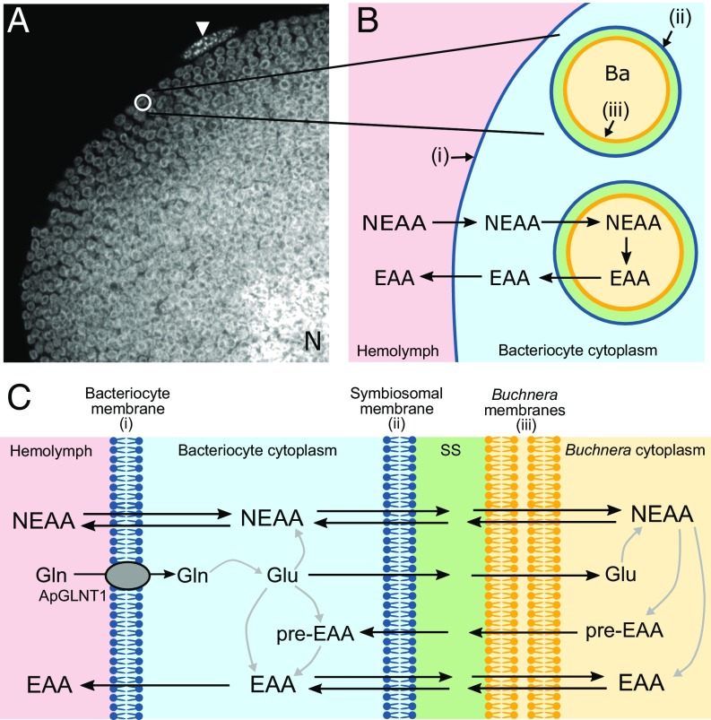
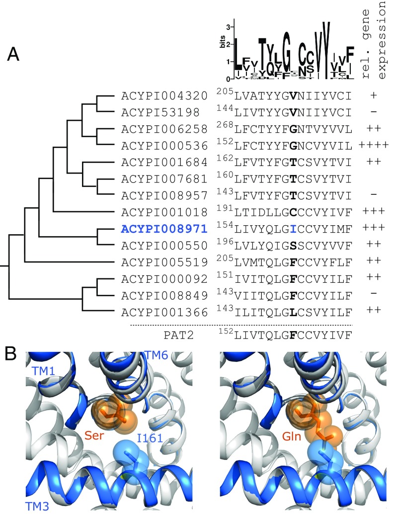
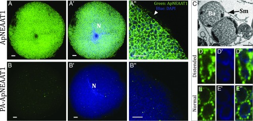
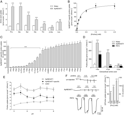
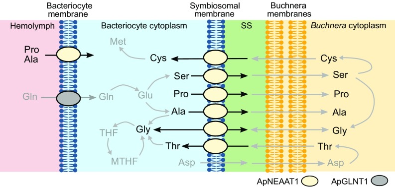
Plant sap-feeding insects are widespread, having evolved to occupy diverse environmental niches despite exclusive feeding on an impoverished diet lacking in essential amino acids and vitamins. Success depends exquisitely on their symbiotic relationships with microbial symbionts housed within specialized eukaryotic bacteriocyte cells. Each bacteriocyte is packed with symbionts that are individually surrounded by a host-derived symbiosomal membrane representing the absolute host-symbiont interface. The symbiosomal membrane must be a dynamic and selectively permeable structure to enable bidirectional and differential movement of essential nutrients, metabolites, and biosynthetic intermediates, vital for growth and survival of host and symbiont. However, despite this crucial role, the molecular basis of membrane transport across the symbiosomal membrane remains unresolved in all bacteriocyte-containing insects. A transport protein was immunolocalized to the symbiosomal membrane separating the pea aphid Acyrthosiphon pisum from its intracellular symbiont Buchnera aphidicola The transporter, A. pisum nonessential amino acid transporter 1, or ApNEAAT1 (gene: ACYPI008971), was characterized functionally following heterologous expression in Xenopus oocytes, and mediates both inward and outward transport of small dipolar amino acids (serine, proline, cysteine, alanine, glycine). Electroneutral ApNEAAT1 transport is driven by amino acid concentration gradients and is not coupled to transmembrane ion gradients. Previous metabolite profiling of hemolymph and bacteriocyte, alongside metabolic pathway analysis in host and symbiont, enable prediction of a physiological role for ApNEAAT1 in bidirectional host-symbiont amino acid transfer, supplying both host and symbiont with indispensable nutrients and biosynthetic precursors to facilitate metabolic complementarity.

Keywords: amino acid transport; metabolic integration; symbiosis.


Conflict of interest statement

The authors declare no conflict of interest.

Figures

Fig. 1.
Fig. 1.
The aphid/Buchnera symbiotic boundary and the role in amino acid exchange. (A) A. pisum bacteriocytes each harbor thousands of bacterial endosymbionts (Buchnera aphidicola). A greyscale confocal (magnification: 630×) image showing DAPI-associated fluorescence (identifying nuclear and Buchnera DNA) through an A. pisum bacteriocyte packed full with Buchnera endosymbionts (visualized by their typical spherical shape, 3 μm in diameter). N represents host nucleus. The arrowhead indicates a sheath cell on the bacteriocyte periphery. (B) Schematic representation of the aphid/Buchnera boundary highlighting the endosymbiotic paradigm, where the host supplies symbiont with NEAAs and the symbiont provides host with EAAs. A series of membranes separate the hemolymph from the symbiont: (i) the aphid (host) bacteriocyte membrane (blue) separates hemolymph from bacteriocyte cytosol; (ii) the host-derived symbiosomal membrane (blue) separates each individual Buchnera from the bacteriocyte cytosol; (iii) the outer and inner membranes (yellow) of the gram-negative Buchnera. Ba, B. aphidicola. (C) More detailed schematic representation of the putative steps in NEAA and EAA transport across the aphid/Buchnera symbiotic boundary. The only identified amino acid transporter to date is the glutamine-selective ApGLNT1, which is localized in the bacteriocyte membrane (28). Much of the glutamine taken into the bacteriocyte is converted into glutamate, which can either be transported across the symbiosomal membrane or converted by bacteriocyte enzymes into NEAAs. The NEAAs must cross the symbiosomal membrane to be utilized by Buchnera and in the Buchnera-mediated production of other NEAAs, EAAs, or EAA precursors (pre-EAA), all of which can exit across the symbiosomal membrane back into the bacteriocyte cytosol. SS, symbiosomal space.
Fig. 2.
Fig. 2.
Identification of aphid ApNEAAT1 (ACYPI008971) as a putative carrier of small NEAAs expressed in the bacteriocyte. (A) Left column: phylogeny showing the relationship between all 14 SLC36-related A. pisum AAAP transporters from the arthropod expanded AAAP clade (30, 31). Phylogenetic tree based on previously published phylogenies (30, 50). Middle column: portion of a full sequence alignment (by PROMALS3D) showing the central section of TM3 with a representation of the variability at each residue position shown above as a Sequence Logo. The residues highlighted in bold are equivalent to both F159 in rat PAT2 (slc36a2) and V104 in LeuT (39, 51). ACYPI008971 has I161 (blue) at this residue position. Right column: Representation of relative gene expression of each transporter within the bacteriocyte structure as a whole. −, not expressed; ++++, most highly expressed; +++, ≤35%; ++, ≤15%, +, ≤1% expression of the most highly expressed amino acid transporter [summary of gene expression determined by RNAseq which is consistent with earlier estimates using qPCR (28, 30)]. ACYPI007681 expression was not determined. (B) A structural model of ACYPI008971 was created using I-TASSER and aligned against the highest-scoring crystal (3L1L of the arginine transporter AdiC, gray). Sections of ACYPI008971 TM1, TM3, and TM6 are shown as blue ribbons. ACYPI008971 I161 (blue sticks and spheres) projects toward the substrate binding pocket. When serine or glutamine (orange sticks and spheres) were positioned in the binding pocket, using the arginine in the 3L1L crystal as a guide, it shows that I161 is likely to limit binding pocket space so that ACYPI008971 may transport amino acids with shorter (Ser) rather than longer (Gln) side-chains.
Fig. 3.
Fig. 3.
Immunolocalization of ApNEAAT1 to the symbiosomal and bacteriocyte membranes of isolated bacteriocyte cells. (A) Immunolocalization of ApNEAAT1 (green) reveals extensive punctate staining around individual Buchnera cells. (A′) Merge of the anti-ApNEAAT1 image and DAPI-stained nuclear and Buchnera DNA (blue). (A″) Magnified region of bacteriocyte cell showing merge of anti-ApNEAAT1 localization (green) and DAPI-stained DNA (blue), arrowhead marks localization to the bacteriocyte cell membrane. (Scale bars, 10 μm.) (BB″) Comparable control experiments were performed with isolated A. pisum bacteriocytes with peptide preadsorbed (PA) anti-ApNEAAT1 antibody. The secondary antibody was Alexa-Fluor 568 donkey anti-rabbit IgG (H+L) (Scale bars, 10 μm). N, bacteriocyte cell nucleus. (C) TEM of distended symbiosomal membrane (Sm) enclosing 2 B. aphidicola (Ba). (Reprinted from ref. , with permission from Elsevier.) Left to right: (D) Immunolocalization of ApNEAAT1 to the distended symbiosomal membrane; (D′) DAPI-stained Buchnera cells; (D″) merge of the anti-ApNEAAT1 image (green) and DAPI-stained Buchnera DNA (blue). (EE″) Comparable images of 2 Buchnera surrounded by their own symbiosomal membranes. For all images, a single representative confocal plane is shown for 3 replicated localization experiments (SI Appendix, Fig. S2).
Fig. 4.
Fig. 4.
The aphid amino acid carrier ApNEAAT1 transports the NEAAs proline, serine, alanine, glycine, and cysteine. (A) Uptake of various radiolabeled amino acids (10 µM) into ApNEAAT1-expressing and water-injected (control) oocytes measured in the absence of extracellular Na+ at pH 5.5. n = 20. ***P < 0.001; **P < 0.01; NS (not significant), P > 0.05 vs. water (2-way, unpaired t tests). (B) ApNEAAT1-specific, concentration-dependent proline uptake. Uptake into water-injected oocytes was subtracted from total uptake. Curve is fitted to Michaelis–Menten kinetics [Km = 179 ± 33 µM; Vmax = 120 ± 6 pmol.oocyte−1.(40 min)−1; r2 = 0.986]. n = 20. (C) Proline uptake in the absence (control) and presence of amino acids or analogs (all 10 mM except Tyr which is 2.5 mM). All are l-isomers unless indicated otherwise. ApNEAAT1-specific uptake is calculated by subtraction of uptake into water-injected oocytes and is expressed as percent control (that in the absence of inhibitor). Tau, taurine. n = 16–20. ***P < 0.001 vs. control; all other bars are P > 0.05 vs. control (1-way ANOVA with Sidak’s posttest). (D) Trans-stimulation of proline ([5 mM]i) efflux via ApNEAAT1 and PAT2 (rat slc36a2) by various extracellular amino acids (10 mM) was measured under Na+-free conditions at extracellular pH 5.5 (10 min). n = 4–5. ***P < 0.001; NS, P > 0.05 vs. water (2-way ANOVA with Tukey’s posttest). (E) Proline uptake in Na+-free conditions over the pH range 5.0–8.0. n = 20. The only significant difference found within each group was in ApNEAAT1-specific uptake: pH 6.5 vs. pH 5.5, P = 0.046 (2-way ANOVA with Tukey’s posttest). (F) Proline-associated inward current in PAT2-expressing but not ApNEAAT1-expressing or uninjected (control) oocytes as measured by 2-electrode voltage clamp. Oocytes were clamped at resting VM (−30 mV), superfused with Na+-free, pH 5.5 buffer and exposed to proline (0.1 to 1 mM). Mean data are shown in (SI Appendix, Table S1) and for ApNEAAT1 in the Inset. (Inset) As a direct comparison, proline uptake via ApNEAAT1 was measured under the same conditions as current measurement. ***P < 0.001.
Fig. 5.
Fig. 5.
A schematic model depicting the proposed physiological function of the amino acid transporter ApNEAAT1 in amino acid transfer across the bacteriocyte and symbiosomal membranes in the aphid/Buchnera bacteriocyte. The predicted pathways are based on the membrane localization and functional characterization of ApNEAAT1 here, alongside metabolite profiling of hemolymph and bacteriocyte, and host and symbiont metabolic pathway analysis (protein and gene expression) in previous investigations (, , , , , –35). ApNEAAT1 substrates are identified in black text with nonsubstrates presented in gray text. The bacteriocyte membrane ApNEAAT1 is depicted as mediating influx into the bacteriocyte. However, if the concentration of any ApNEAAT1 substrate within the bacteriocyte was greater than in the hemolymph, it could also mediate efflux from bacteriocyte to hemolymph to supply other tissues, for example, during growth and development. MTHF, 5,10-methylene tetrahydrofolate; SS, symbiosomal space.

References

    1. Douglas A. E., The Symbiotic Habit (Princeton University Press, 2010).
    1. DeSalle R., Perkins S. L., Welcome to the Microbiome: Getting to Know the Trillions of Bacteria and Other Microbes in, on, and Around You (Yale University Press, 2015).
    1. Yong E., I Contain Multitudes: The Microbes within Us and a Grander View of Life (HarperCollins, 2016).
    1. Douglas A. E., Multiorganismal insects: Diversity and function of resident microorganisms. Annu. Rev. Entomol. 60, 17–34 (2015). - PMC - PubMed
    1. Moran N. A., McCutcheon J. P., Nakabachi A., Genomics and evolution of heritable bacterial symbionts. Annu. Rev. Genet. 42, 165–190 (2008). - PubMed

Publication types

LinkOut - more resources