

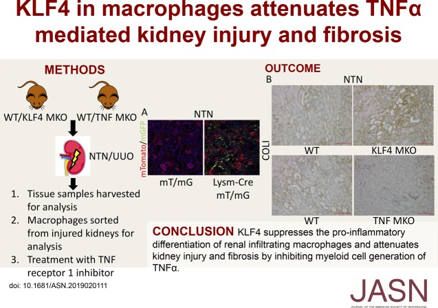
KLF4 in Macrophages Attenuates TNF α-Mediated Kidney Injury and Fibrosis
- PMID: 31337692
- PMCID: PMC6779357
- DOI: 10.1681/ASN.2019020111
KLF4 in Macrophages Attenuates TNF α-Mediated Kidney Injury and Fibrosis
Abstract
Background: Polarized macrophage populations can orchestrate both inflammation of the kidney and tissue repair during CKD. Proinflammatory M1 macrophages initiate kidney injury, but mechanisms through which persistent M1-dependent kidney damage culminates in fibrosis require elucidation. Krüppel-like factor 4 (KLF4), a zinc-finger transcription factor that suppresses inflammatory signals, is an essential regulator of macrophage polarization in adipose tissues, but the effect of myeloid KLF4 on CKD progression is unknown.
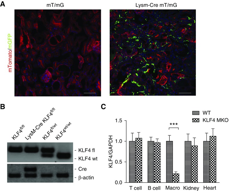
Methods: We used conditional mutant mice lacking KLF4 or TNFα (KLF4's downstream effector) selectively in myeloid cells to investigate macrophage KLF4's role in modulating CKD progression in two models of CKD that feature robust macrophage accumulation, nephrotoxic serum nephritis, and unilateral ureteral obstruction.
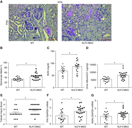
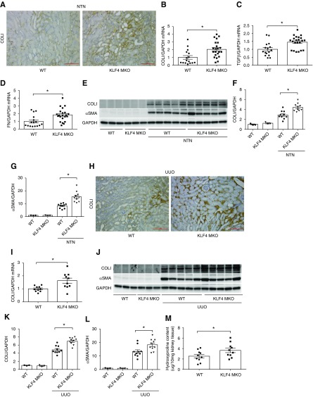
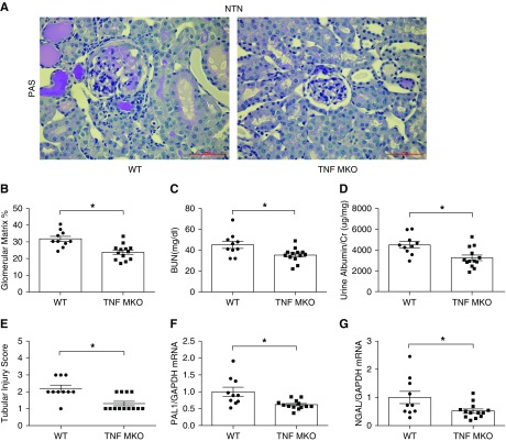
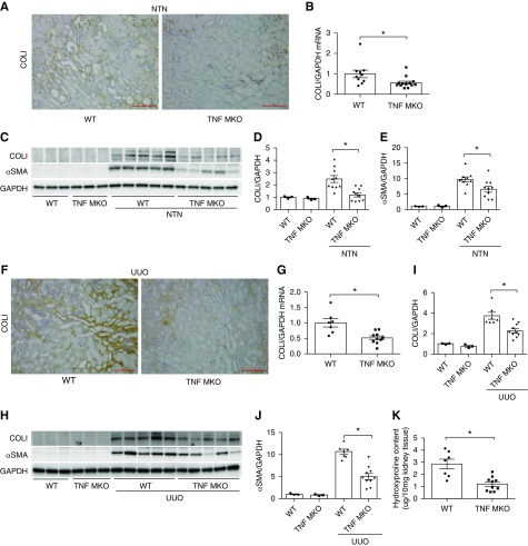
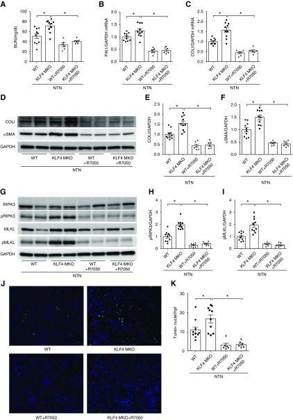
Results: In these murine CKD models, KLF4 deficiency in macrophages infiltrating the kidney augmented their M1 polarization and exacerbated glomerular matrix deposition and tubular epithelial damage. During the induced injury in these models, macrophage-specific KLF4 deletion also exacerbated kidney fibrosis, with increased levels of collagen 1 and α-smooth muscle actin in the injured kidney. CD11b+Ly6Chi myeloid cells isolated from injured kidneys expressed higher levels of TNFα mRNA versus wild-type controls. In turn, mice bearing macrophage-specific deletion of TNFα exhibited decreased glomerular and tubular damage and attenuated kidney fibrosis in the models. Moreover, treatment with the TNF receptor-1 inhibitor R-7050 during nephrotoxic serum nephritis reduced damage, fibrosis, and necroptosis in wild-type mice and mice with KLF4-deficient macrophages, and abrogated the differences between the two groups in these parameters.
Conclusions: These data indicate that macrophage KLF4 ameliorates CKD by mitigating TNF-dependent injury and fibrosis.
Keywords: KLF4; macrophages; necroptosis; renal fibrosis.
Copyright © 2019 by the American Society of Nephrology.
Figures

References
-
- Grivennikov SI, Tumanov AV, Liepinsh DJ, Kruglov AA, Marakusha BI, Shakhov AN, et al.: Distinct and nonredundant in vivo functions of TNF produced by t cells and macrophages/neutrophils: Protective and deleterious effects. Immunity 22: 93–104, 2005 - PubMed
MeSH terms
Substances
Grants and funding
LinkOut - more resources
Full Text Sources
Medical
Research Materials
