. 2019 Jul 23;10(4):e01065-19.
doi: 10.1128/mBio.01065-19.

Stable Occupancy of the Crimean-Congo Hemorrhagic Fever Virus-Encoded Deubiquitinase Blocks Viral Infection

Affiliations

Stable Occupancy of the Crimean-Congo Hemorrhagic Fever Virus-Encoded Deubiquitinase Blocks Viral Infection

Florine E M Scholte et al. mBio. .

Abstract

Crimean-Congo hemorrhagic fever virus (CCHFV) infection can result in a severe hemorrhagic syndrome for which there are no antiviral interventions available to date. Certain RNA viruses, such as CCHFV, encode cysteine proteases of the ovarian tumor (OTU) family that antagonize interferon (IFN) production by deconjugating ubiquitin (Ub). The OTU of CCHFV, a negative-strand RNA virus, is dispensable for replication of the viral genome, despite being part of the large viral RNA polymerase. Here, we show that mutations that prevent binding of the OTU to cellular ubiquitin are required for the generation of recombinant CCHFV containing a mutated catalytic cysteine. Similarly, the high-affinity binding of a synthetic ubiquitin variant (UbV-CC4) to CCHFV OTU strongly inhibits viral growth. UbV-CC4 inhibits CCHFV infection even in the absence of intact IFN signaling, suggesting that its antiviral activity is not due to blocking the OTU's immunosuppressive function. Instead, the prolonged occupancy of the OTU with UbV-CC4 directly targets viral replication by interfering with CCHFV RNA synthesis. Together, our data provide mechanistic details supporting the development of antivirals targeting viral OTUs.IMPORTANCE Crimean-Congo hemorrhagic fever virus is an important human pathogen with a wide global distribution for which no therapeutic interventions are available. CCHFV encodes a cysteine protease belonging to the ovarian tumor (OTU) family which is involved in host immune suppression. Here we demonstrate that artificially prolonged binding of the OTU to a substrate inhibits virus infection. This provides novel insights into CCHFV OTU function during the viral replicative cycle and highlights the OTU as a potential antiviral target.

Keywords: Nairoviridae; RNA replication; antiviral agents; bunyavirus; innate immunity; interferon-stimulated gene-15; proteases; ubiquitination.


Figures

FIG 1
FIG 1
Generation of recombinant CCHFV containing an inactive OTU. (a) Recovery of ZsG-encoding CCHFV reporter viruses containing OTU mutations. Huh7 cells were transfected with plasmids encoding the full-length S, M, and L genome segments and with helper plasmids encoding NP, codon-optimized L, and T7 RNA polymerase. After 5 days, supernatants were transferred to fresh Huh7 cells, and ZsG fluorescence was imaged to determine successful virus rescue. WT, wild type. (b to d) Growth kinetics of recombinant CCHFV-ZsG viruses containing the indicated OTU mutations in A549, BSR-T7/5, and A549 RIG-I KO cells (multiplicity of infection [MOI] of 0.1). At the indicated time points (days [D]), ZsG fluorescence was quantified as a measure of viral replication. Data are presented as means ± standard deviations (SD) of results from three replicates.
FIG 2
FIG 2
Lethality of CCHFV OTU mutants in an IFNAR−/− mouse model. Female IFNAR−/− mice (6 weeks of age) were infected subcutaneously with 100 TCID50 of recombinant CCHFV (8 mice per group) and analyzed for weight (a) and survival (b). Animals were monitored daily for clinical signs of illness. Animal weights are shown as means ± SD and represent percentages of weight changes relative to a baseline set at 1 day before challenge. (c) The presence of viral RNA in blood and various homogenized tissues was analyzed using quantitative RT-PCR. The two survivors are indicated with open symbols. ***, statistical significance (P < 0.001) of results of comparisons between CCHFV-wt and CCHFV-C40A/Q16R.
FIG 3
FIG 3
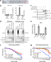
UbV-CC4 blocks CCHFV OTU activity. (a) Alignment of the C-terminal sequences of Ub and the CC4 and AA Ub variants. Residue variations are highlighted in red. (b) Analysis of OTU (wt and C40A) interactions with HA-Ub or UbV-CC4 using TR-FRET. HEK293T cells were cotransfected with plasmids expressing V5-OTU and HA-Ub or plasmids expressing V5-OTU and FLAG-UbV-CC4. A plasmid expressing GFP was used as a control. After 2 days, samples were harvested and incubated with donor and acceptor fluorophores. Protein-protein proximity was determined by measuring the intensity of the fluorescent signal. Data are presented as means ± SD of results from three replicates. (c) To confirm the interaction of OTU-wt with UbV-CC4, HEK293T cells were transfected with plasmids expressing HA-OTU-wt and FLAG-UbV (FLAG-UbV-AA or FLAG-UbV-CC4). Lysates were immunoprecipitated (IP) with HA antibodies and analyzed by Western blotting (WB). (d) To assess whether UbV-CC4 affects CCHFV OTU activity, Huh7 cells were cotransfected with plasmids encoding HA-Ub (DUB assay) or the proteins required for ISGylation (V5-ISG15, Ube1L, UbcH8, and HERC5). After 48 h, levels of protein ubiquitination and ISGylation were assessed by Western blotting. (e) To assess the effect of UbV-CC4 on OTU-mediated immune suppression in an overexpression assay, Huh7 cells were cotransfected with a reporter plasmid encoding firefly luciferase under the control of the IFN-β promoter, and a plasmid encoding constitutively active RIG-I CARD. After 48 h, luciferase activity was determined as a measure of RIG-I-mediated IFN-β expression. A plasmid expressing Renilla luciferase was used for normalization. Data are presented as means ± SD of results from four replicates. (f and g) In vitro analysis of the effect of UbV-CC4 on OTUs from different CCHFV strains (f) or other nairovirus species (g). Reactions to determine the inhibitory activity of Ub-CC4 against the different OTUs were performed using inhibitor concentrations ranging from 3.9 nM to 5 μM. OTUs were incubated with the inhibitor for 2 min, and then the reactions were initiated by adding Ub-AMC and were monitored by quantification of the increase in fluorescence. Reaction rates were determined using the linear portion of the curves, and percent inhibition was calculated. IC50 and related errors were determined in the SigmaPlot 12 enzyme kinetics module utilizing Michaelis-Menten kinetics (Systat Software, Inc.). Data are presented as means ± SD of results from three replicates.
FIG 4
FIG 4
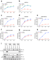
UbV-CC4 can block CCHFV replication in the absence of RIG-I-mediated immune responses. (a and b) To determine the effect of UbV-CC4 on the release of progeny virus, A549 cells stably expressing UbV-AA (A549-AA) or UbV-CC4 (A549-CC4) were infected with either the ZsG reporter virus (CCHFV-ZsG-wt) (a) or the unlabeled parental CCHFV (CCHFV-wt) (b) at an MOI of 0.01. Infectious titers were determined at the indicated time points. Data are presented as means ± SD of results from three independent experiments performed in triplicate. (c) To assess the effect of UbV-CC4 on viral replication, A549 cells stably expressing UbV-AA or UbV-CC4 were infected with a CCHFV reporter virus (CCHFV-ZsG; MOI of 0.1). (d) LASV-ZsG (MOI of 0.1) was used as a control. ZsG reporter activity was quantified at the indicated time points as a measure of viral replication. Data are presented as means ± SD of results from three replicates. (e) To determine whether RIG-I signaling is involved in the CC4-mediated suppression of CCHFV replication, A549 RIG-I KO cells stably expressing UbV-AA or UbV-CC4 were infected with CCHFV-ZsG (MOI of 0.1) and ZsG reporter activity was quantified at the indicated time points. Data are presented as means ± SD of results from three replicates. (f to h) To confirm that direct binding of the UbV-CC4 to the CCHFV OTU is responsible for the inhibition of CCHFV replication, we employed recombinant viruses containing OTU mutations that disrupt binding to Ub (Q16R) or to ISG15 (P77D) or to both Ub and ISG15 (A129R). A549 cells stably expressing UbV were infected with these CCHFV OTU mutants (MOI of 0.1), and ZsG fluorescence was determined as a measure of viral replication at the indicated time points. Data are presented as means ± SD of results from three replicates. (i) To confirm the interaction of the OTU (mutants) with UbV-CC4, we performed coimmunoprecipitation assays. HEK293T cells were transfected with the indicated plasmids, and cell lysates were harvested after 2 days.
FIG 5
FIG 5
UbV-CC4 inhibits viral RNA transcription and/or replication. (a) VLPs were used to determine whether UbV-CC4 affects CCHFV entry. VLPs were generated by transfecting Huh7 cells with plasmids encoding CCHFV NP, GPC, codon-optimized L, T7 polymerase, and a NanoLuc (Nluc)-encoding minigenome. VLP-containing supernatants were harvested after 3 days and transferred to A549 cells stably expressing UbV-AA or UbV-CC4. NanoLuc activity was quantified the next day as a measure of VLP activity. Data are presented as means ± SD of results from four replicates. RLU, relative light units. (b) To analyze if UbV-CC4 affects early replication steps after entry, we used a NanoLuc-expressing reporter virus (CCHFV-Nluc). A549 cells stably expressing UbV were infected (MOI of 5), and NanoLuc activity was determined at the indicated time points. Data are presented as means ± SD of results from three replicates. (c) A minigenome assay was used to determine if UbV-CC4 directly affects CCHFV genome amplification. Huh7 cells were cotransfected with plasmids encoding CCHFV NP, codon-optimized L, T7 polymerase, and a NanoLuc-encoding minigenome. After 2 days, NanoLuc activity was quantified as a measure of CCHFV RdRp activity. Constitutively active Renilla luciferase was used as a transfection control. Data are presented as means ± SD of results from three replicates. (d) To analyze CCHFV minigenome RNA levels, RNA was isolated from Huh7 cells transfected with the minigenome components described for panel c. Minigenome RNA levels were quantified using qRT-PCR. Data are presented as means ± SD of results from six replicates. (e) To investigate if UbV-CC4 binding to the OTU interferes with L-NP interaction, HEK293T cells were transfected with plasmids expressing CCHFV NP, codon-optimized V5-L, and UbV-AA or UbV-CC4. L-NP interaction was assessed by coimmunoprecipitating V5-L and determining the associated NP levels.
FIG 6
FIG 6
Schematic model of OTU stable interaction with cellular Ub or UbV-CC4 leading to the block of viral RNA replication. (Panel I) Wild-type OTU possesses deubiquitinase activity, which allows the deconjugation of Ub chains. (Panel II) Catalytically inactive OTU (C40A) can engage ubiquitinated substrates but is not able to cleave Ub chains. The formation of a Ub-OTU complex interferes with viral replication. (Panel III) Adding an additional mutation that prevents binding to Ub (Q16R) to catalytically inactive OTU (C40A) disrupts the formation of Ub-OTU complexes and restores viral replication. (Panel IV) Binding of a synthetic Ub variant (UbV-CC4) to wild-type OTU interferes with viral replication.

Similar articles

Cited by

References

    1. Spengler JR, Bergeron É, Rollin PE. 2016. Seroepidemiological studies of Crimean-Congo hemorrhagic fever virus in domestic and wild animals. PLoS Negl Trop Dis 10:1–28. doi:10.1371/journal.pntd.0004210. - DOI - PMC - PubMed
    1. Spengler JR, Bergeron E, Spiropoulou CF. 2019. Crimean-Congo hemorrhagic fever and expansion from endemic regions. Curr Opin Virol 34:70–78. doi:10.1016/j.coviro.2018.12.002. - DOI - PMC - PubMed
    1. Estrada-Peña A, Ruiz-Fons F, Acevedo P, Gortazar C, de la Fuente J. 2013. Factors driving the circulation and possible expansion of Crimean-Congo haemorrhagic fever virus in the western Palearctic. J Appl Microbiol 114:278–286. doi:10.1111/jam.12039. - DOI - PubMed
    1. García Rada A. 2016. First outbreak of Crimean-Congo haemorrhagic fever in Western Europe kills one man in Spain. BMJ 4891:i4891. - PubMed
    1. Negredo A, de la Calle-Prieto F, Palencia-Herrejón E, Mora-Rillo M, Astray-Mochales J, Sánchez-Seco MP, Bermejo Lopez E, Menárguez J, Fernández-Cruz A, Sánchez-Artola B, Keough-Delgado E, Ramírez de Arellano E, Lasala F, Milla J, Fraile JL, Ordobás Gavín M, Martinez de la Gándara A, López Perez L, Diaz-Diaz D, López-García MA, Delgado-Jimenez P, Martín-Quirós A, Trigo E, Figueira JC, Manzanares J, Rodriguez-Baena E, Garcia-Comas L, Rodríguez-Fraga O, García-Arenzana N, Fernández-Díaz MV, Cornejo VM, Emmerich P, Schmidt-Chanasit J, Arribas JR. 2017. Autochthonous Crimean-Congo hemorrhagic fever in Spain. N Engl J Med 377:154–161. doi:10.1056/NEJMoa1615162. - DOI - PubMed

Publication types

MeSH terms