. 2019 Jul 23;9(1):10619.
doi: 10.1038/s41598-019-46831-3.

Genomic and molecular characterisation of Escherichia marmotae from wild rodents in Qinghai-Tibet plateau as a potential pathogen

Affiliations

Genomic and molecular characterisation of Escherichia marmotae from wild rodents in Qinghai-Tibet plateau as a potential pathogen

Sha Liu et al. Sci Rep. .

Abstract

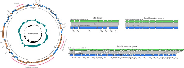
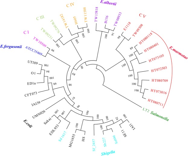
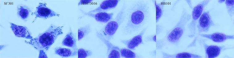
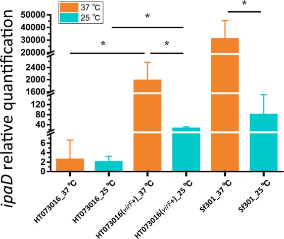
Wildlife is a reservoir of emerging infectious diseases of humans and domestic animals. Marmota himalayana mainly resides 2800-4000 m above sea level in the Qinghai-Tibetan Plateau, and is the primary animal reservoir of plague pathogen Yersinia pestis. Recently we isolated a new species, Escherichia marmotae from the faeces of M. himalayana. In this study we characterised E. marmotae by genomic analysis and in vitro virulence testing to determine its potential as a human pathogen. We sequenced the genomes of the seven E. marmotae strains and found that they contained a plasmid that carried a Shigella-like type III secretion system (T3SS) and their effectors, and shared the same O antigen gene cluster as Shigella dysenterae 8 and E. coli O38. We also showed that E. marmotae was invasive to HEp-2 cells although it was much less invasive than Shigella. Thus E. marmotae is likely to be an invasive pathogen. However, E. marmotae has a truncated IpaA invasin, and lacks the environmental response regulator VirF and the IcsA-actin based intracellular motility, rendering it far less invasive in comparison to Shigella. E. marmotae also carried a diverse set of virulence factors in addition to the T3SS, including an IS1414 encoded enterotoxin gene astA with 37 copies, E. coli virulence genes lifA/efa, cif, and epeA, and the sfp gene cluster, Yersinia T3SS effector yopJ, one Type II secretion system and two Type VI secretion systems. Therefore, E. marmotae is a potential invasive pathogen.


Conflict of interest statement

The authors declare no competing interests.

Figures

Figure 1
Figure 1
Circular representations of the pEM148 plasmid of E. marmotae. From the outside in (to scale): circle 1 represents genes on the positive and negative strands (scale is marked in 50 kb), circle 2 shows a plot of GC content (higher values outward), and circle 3 shows a plot of GC skew (G − C)/(G + C). The red curves indicate the region to be compared. Arrows of inset indicate predicted ORFs in both strands. Shown below gene bar are locus tags. Regions in light gray indicate homologous sequences and percentages of identity between two homologous genes at the nucleotide level. The inset depicts the comparisons of the plasmid regions of T3SS, T2SS and sfp gene cluster with the corresponding regions of pCP301 of Shigella flexneri str. 301 (NC_004851), E. coli 545 chromosome (NZ_CP018976) and sfp cluster of E. coli plasmid pCFSAN004177G_03(CP012494).
Figure 2
Figure 2
Phylogenetic tree of E. marmotae and 30 representative genomes. The tree was constructed using the maximum likelihood algorithms in Phylip based on the core genome SNPs. Escherichia clade I–V are marked as C I to C V. C V belonged to the species E. marmotae. Values on the branch are bootstrap values out of 100 from 1000 replicates. Species and strain names are colour coded. Note that Shigella strains belong to E. coli. Salmonella Typhimurium strain LT2 is used as an outgroup.
Figure 3
Figure 3
Invasion of epithelial cells by E. marmotae in vitro. HEp-2 cell invasion by E. marmotae HT073016, S. flexneri str. 301 (positive control) and E. coli HB101 (negative control) as labelled. Infected HEp-2 cells were fixed by methanol and then stained with Geimsa.
Figure 4
Figure 4
Expression of ipaD under different temperature conditions as determined by qRT-PCR. Total RNA was harvested from HT073016, HT073016(virF+) and S. flexneri strain 301 cultivated at 25 °C and 37 °C. Relative transcript levels were calculated using the ∆∆CT method and fold changes in comparison to HT073016 at 37 °C. All values have been normalized to the endogenous reference gene recA. Means and standard deviations stand for three independent experiments were shown with * being p < 0.05 in the indicated comparisons. Error bars are +SD.

References

    1. Daszak P, Cunningham AA, Hyatt AD. Emerging Infectious Diseases of Wildlife–Threats to Biodiversity and Human Health. Science. 2000;287:443. doi: 10.1126/science.287.5452.443. - DOI - PubMed
    1. Neumann G, Chen H, Gao G, Shu Y, Kawaoka Y. H5N1 influenza viruses: outbreaks and biological properties. Cell Res. 2010;20:51–61. doi: 10.1038/cr.2009.124. - DOI - PMC - PubMed
    1. Burgdorfer W. Discovery of the Lyme disease spirochete and its relation to tick vectors. The Yale journal of biology and medicine. 1984;57:515–520. - PMC - PubMed
    1. Steppan S, et al. Molecular phylogeny of the marmots (Rodentia: Sciuridae): tests of evolutionary and biogeographic hypotheses. Syst Biol. 1999;48:715–734. doi: 10.1080/106351599259988. - DOI - PubMed
    1. Qian, Q. et al. Mapping risk of plague in Qinghai-Tibetan Plateau, China. BMC Infect Dis. 14, 10.1186/1471-2334-14-382 (2014). - PMC - PubMed

Publication types

Substances

Supplementary concepts