. 2019 Jul 23;9(1):10672.
doi: 10.1038/s41598-019-46958-3.

A collagen-based microwell migration assay to study NK-target cell interactions

Affiliations

A collagen-based microwell migration assay to study NK-target cell interactions

Per E Olofsson et al. Sci Rep. .

Abstract

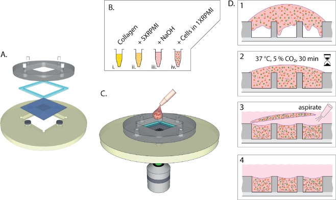
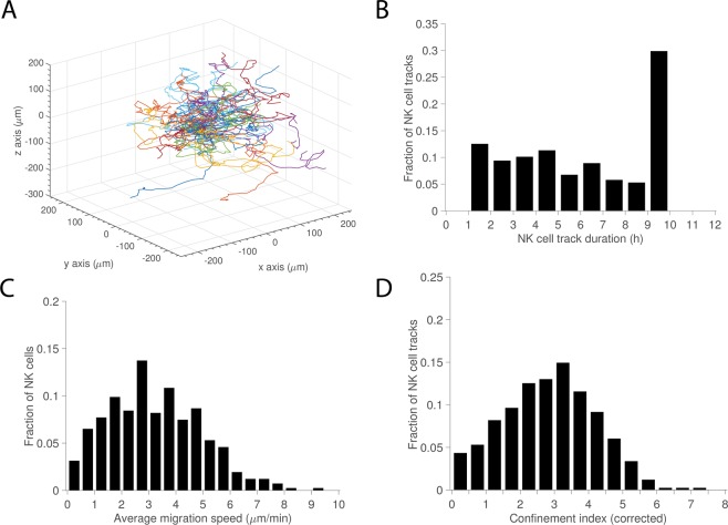
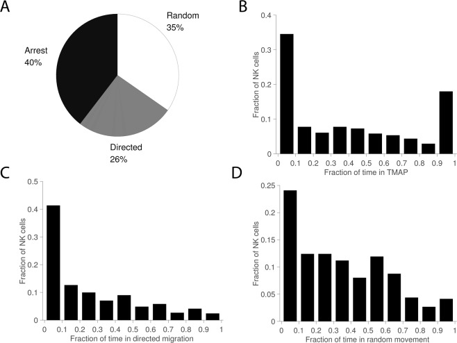
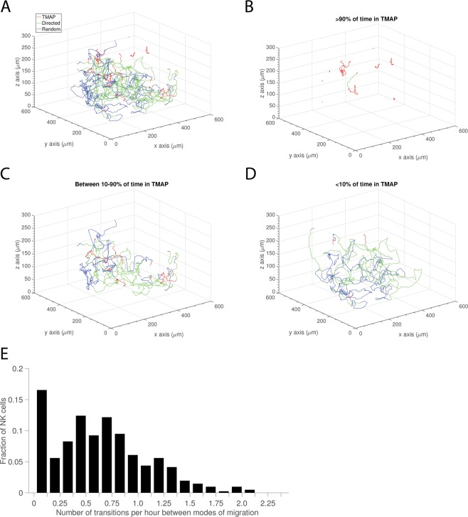
Natural killer (NK) cell cytotoxicity in tissue is dependent on the ability of NK cells to migrate through the extracellular matrix (ECM) microenvironment. Traditional imaging studies of NK cell migration and cytotoxicity have utilized 2D surfaces, which do not properly reproduce the structural and mechanical cues that shape the migratory response of NK cells in vivo. Here, we have combined a microwell assay that allows long-term imaging and tracking of small, well-defined populations of NK cells with an interstitial ECM-like matrix. The assay allows for long-term imaging of NK-target cell interactions within a confined 3D volume. We found marked differences in motility between individual cells with a small fraction of the cells moving slowly and being confined to a small volume within the matrix, while other cells moved more freely. A majority of NK cells also exhibited transient variation in their motility, alternating between periods of migration arrest and movement. The assay could be used as a complement to in vivo imaging to study human NK cell heterogeneity in migration and cytotoxicity.


Conflict of interest statement

The authors declare no competing interests.

Figures

Figure 1
Figure 1
Schematic figure of experimental setup and hydrogel embedding procedure. (A) Exploded view of the microchip platform consisting of plastic holder with embedded stainless-steel discs, microchip, gasket, and plastic lid with embedded magnets (B) Procedure for preparing collagen-embedded cells mixtures. Stock solution of collagen monomers dissolved in acetic acid (i) was brought to the right concentration by addition of concentrated cell medium (ii) and reconstituted by adding NaOH (iii) to which a mixture of NK cells and target cells suspended in RPMI was added (iv). (C) The cell-collagen mix was rapidly deposited onto the microwell chip inserted in the assembled holder. (D) Schematic view of the deposit and maturation of the collagen matrix in the microwells. The viscous collagen-cell mixture was poured into the wells (1) and then incubated under physiological conditions for 30 min (2). When the matrix had set, cell medium was gently streamed over the wells which caused excess matrix to detach from the chip (3) leaving only cell-collagen mixture in the wells (4).
Figure 2
Figure 2
NK cell migration in 3D. (A) Track plot of NK cell migration trajectories centered at their respective starting points. (B) Distribution of NK cell track durations (>1 h). (C) Distribution of average migration speeds calculated from individual tracks. (D) Corrected confinement index calculated from individual tracks. Total number of NK cells n = 411.
Figure 3
Figure 3
Mode of migration. (A) Pie chart showing distribution of time spent in transient migration arrest (TMAP), random movement, and directed migration. (B) Distribution of fractions of time spent in TMAP for individual tracks. (C) Distributions of fractions of time spent in directed migration. (D) Distribution of fractions of time spent in random movement. Total number of NK cells n = 411.
Figure 4
Figure 4
Transient migration behavior. (A) Track plot of NK cell migration trajectories recorded in a single well divided into modes of migration (n = 25). (B) Track plot of NK cells spending >90% of time in TMAP (n = 10). (C) Track plot of NK cells spending between 90% and 10% of time in TMAP (n = 9). (D) Track plot of NK cells spending < 10% of time in TMAP (n = 6). (E) Distribution of number of transitions between different modes of migration per hour (n = 411).
Figure 5
Figure 5
NK cell-mediated lysis of target cells in 3D collagen matrix. (A) Time-lapse sequence showing a NK cell migrating and killing two target cells. Scale bar = 15 μm. (B) Number of target cell contacts and kills made by individual NK cells. (C) Distribution of duration of contact periods (n = 86). (D) Distribution of duration of contact periods divided by cytolytic and non-cytolytic interactions (ncytolytic = 49 and nnon-cytolytic = 37).

References

    1. Davis DM, et al. The human natural killer cell immune synapse. Proc Natl Acad Sci USA. 1999;96(26):15062–7. doi: 10.1073/pnas.96.26.15062. - DOI - PMC - PubMed
    1. Ljunggren HG, Karre K. In search of the ‘missing self’: MHC molecules and NK cell recognition. Immunol Today. 1990;11(7):237–44. doi: 10.1016/0167-5699(90)90097-S. - DOI - PubMed
    1. Krzewski K, Coligan JE. Human NK cell lytic granules and regulation of their exocytosis. Front Immunol. 2012;3:335. doi: 10.3389/fimmu.2012.00335. - DOI - PMC - PubMed
    1. Kalluri R, Weinberg RA. The basics of epithelial-mesenchymal transition. J Clin Invest. 2009;119(6):1420–8. doi: 10.1172/JCI39104. - DOI - PMC - PubMed
    1. Hynes RO, Naba A. Overview of the matrisome–an inventory of extracellular matrix constituents and functions. Cold Spring Harb Perspect Biol. 2012;4(1):a004903. doi: 10.1101/cshperspect.a004903. - DOI - PMC - PubMed

Publication types

MeSH terms