. 2019 Jul 23;9(1):10611.
doi: 10.1038/s41598-019-47027-5.

Transcriptomics of a KDELR1 knockout cell line reveals modulated cell adhesion properties

Affiliations

Transcriptomics of a KDELR1 knockout cell line reveals modulated cell adhesion properties

Andrea Blum et al. Sci Rep. .

Abstract

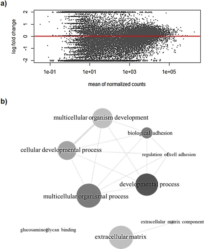
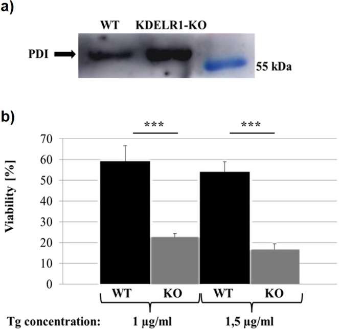
KDEL receptors (KDELRs) represent transmembrane proteins of the secretory pathway which regulate the retention of soluble ER-residents as well as retrograde and anterograde vesicle trafficking. In addition, KDELRs are involved in the regulation of cellular stress response and ECM degradation. For a deeper insight into KDELR1 specific functions, we characterised a KDELR1-KO cell line (HAP1) through whole transcriptome analysis by comparing KDELR1-KO cells with its respective HAP1 wild-type. Our data indicate more than 300 significantly and differentially expressed genes whose gene products are mainly involved in developmental processes such as cell adhesion and ECM composition, pointing out to severe cellular disorders due to a loss of KDELR1. Impaired adhesion capacity of KDELR1-KO cells was further demonstrated through in vitro adhesion assays, while collagen- and/or laminin-coating nearly doubled the adhesion property of KDELR1-KO cells compared to wild-type, confirming a transcriptional adaptation to improve or restore the cellular adhesion capability. Perturbations within the secretory pathway were verified by an increased secretion of ER-resident PDI and decreased cell viability under ER stress conditions, suggesting KDELR1-KO cells to be severely impaired in maintaining cellular homeostasis.


Conflict of interest statement

The authors declare no competing interests.

Figures

Figure 1
Figure 1
Confirmation of KDELR1-KO in HAP1 cells. (a) Sequencing result for the genomic KDELR1 DNA sequence of KDELR1-KO cells (third lane) and wild-type (fourth lane). KDELR1-KO cells show a deletion of „T16“ within the first exon of the KDELR1 gene in HAP1 cells, leading to a premature stop-codon in exon 2 (T188 G189 A190). (b) mRNA level of KDELR1 in wild-type (WT) and KDELR1-KO (KO) HAP1 cells. Represented are the mean values of the TPM (transcripts per million) from poly-A enriched RNA seq (triplicates with standard deviation).
Figure 2
Figure 2
KDELR1-KO affects the transcriptome of HAP1 cells. (a) MA plot representing the difference between wild-type and KDELR1-KO samples. (b) Visualisation of significantly enriched GO terms (adjusted p-value < 0.01) in KDELR1-KO cells compared to HAP1 wild-type. GO terms and their adjusted p-values were used in REViGO to generate three interactive graphs (allowed similarity: medium (0.7), database: Homo sapiens, semantic similarity measure: SimRel), which are shown in one image. The size of the circles represents the frequency of the respective GO term in the gene set. More intensive grey shades were used to show higher significance in the GO analysis. Similar GO terms were connected with lines, whereby the width of each line demonstrates the level of similarity.
Figure 3
Figure 3
KDELR1-KO cells show enhanced migration/proliferation ability but impaired adhesion properties on uncoated surfaces which can be rescued by coating with collagen or laminin. (a) Scratch assay to compare migration behaviour of KDELR1-KO cells to HAP1 wild-type. Areas at time point 0 h were measured and calculated at 100%, scratch areas after 24 h and 48 h incubation in FCS-free medium were measured and presented in a diagram as mean value from 6 replicates. Error bars indicate the corresponding standard deviation, statistical significance was determined by students t-test and significant differences were marked by an asterisks (p < 0.05). Additionally, two-way ANOVA was performed which identified significant differences for samples (p = 0.047) and interaction (p = 0.009). (b) Adhesion assay to compare adhesion behaviour of KDELR1-KO to HAP1 wild-type cells in collagen-coated, laminin-coated or uncoated 96-well plates. Unbound cells were removed by washing with 1 × PBS. After 4 h adhesion time, adherent cells were fixed with 2% PFA, washed twice with 1 × PBS and stained with crystal violet. Absorption was measured at 590 nm after 5 washing steps with sterile water. Diagrams represent the mean value from 15 replicates and the respective standard deviation. Statistical significance was determined by students t-test and significant differences were marked by three asterisks (p < 0.01). Two-way ANOVA additionally indicated significant differences for the samples (p < 0.01), condition (p < 0.01), and interaction (p < 0.01).
Figure 4
Figure 4
KDELR1-KO cells show an increased secretion of PDI and reduced cell viability under ER stress conditions. (a) Secretion of PDI in HAP1 wild-type (WT) and KDELR1-KO cells. In each case, a cell-free culture supernatant was dialysed against PBS, concentrated by VivaSpin centrifugation, and the total protein amount was determined and equally adjusted. For both samples, the same amount of protein was separated by SDS-PAGE and PDI signal intensity was subsequently analysed by western blotting. (b) Sensitivity of HAP1 WT and KDELR1-KO cells under ER stress conditions. Cells were treated with thapsigargin (Tg) for 24 h to induce ER stress and cell viability was analysed via MTT assays. Viability in DMSO control samples was calculated as 100%, viability of Tg-treated cells was determined and is represented in the diagram as mean value from 20 (1 µg/ml Tg) or 10 (1,5 µg/ml Tg) replicates together with the respective standard deviation. Statistical significance was determined by students t-test and significant differences were marked by three asterisks (p < 0.01). Additionally, two-way ANOVA was performed which indicated significant differences for the samples (p < 0.01), condition (p < 0.01), and interaction (p < 0.01).

Similar articles

Cited by

References

    1. Semenza JC, Hardwick KG, Dean N, Pelham HRB. ERD2, a yeast gene required for the receptor-mediated retrieval of luminal ER proteins from the secretory pathway. Cell. 1990;61(7):1349–1357. doi: 10.1016/0092-8674(90)90698-E. - DOI - PubMed
    1. Lewis MJ, Pelham HRB. A human homologue of the yeast HDEL receptor. Nature. 1990;348(6297):162–163. doi: 10.1038/348162a0. - DOI - PubMed
    1. Townsley FM, Wilson DW, Pelham HR. Mutational analysis of the human KDEL receptor: distinct structural requirements for Golgi retention, ligand binding and retrograde transport. EMBO J. 1993;12(7):2821–9. doi: 10.1002/j.1460-2075.1993.tb05943.x. - DOI - PMC - PubMed
    1. Aoe T, et al. The KDEL receptor, ERD2, regulates intracellular traffic by recruiting a GTPase-activating protein for ARF1. EMBO J. 1997;16(24):7305–7316. doi: 10.1093/emboj/16.24.7305. - DOI - PMC - PubMed
    1. Cukierman E, Huber I, Rotman M, Cassel D. The ARF1 GTPase-activating protein: zinc finger motif and Golgi complex localization. Science. 1995;270(5244):1999–2002. doi: 10.1126/science.270.5244.1999. - DOI - PubMed

Publication types