. 2020 Mar;27(3):1023-1035.
doi: 10.1038/s41418-019-0395-5. Epub 2019 Jul 23.

LZTR1 facilitates polyubiquitination and degradation of RAS-GTPases

Affiliations

LZTR1 facilitates polyubiquitination and degradation of RAS-GTPases

Taiki Abe et al. Cell Death Differ. 2020 Mar.

Abstract

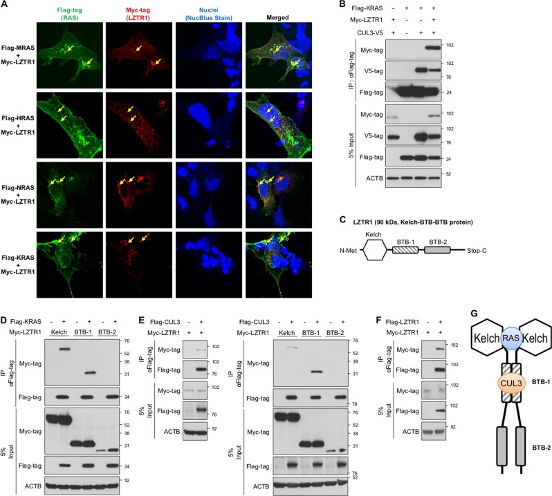
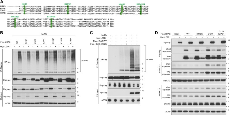
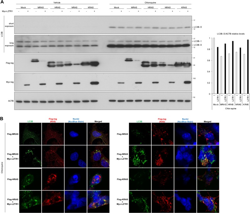
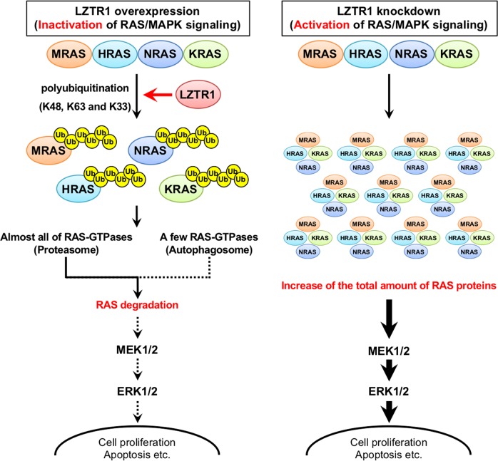
Leucine zipper-like transcriptional regulator 1 (LZTR1) encodes a member of the BTB-Kelch superfamily, which interacts with the Cullin3 (CUL3)-based E3 ubiquitin ligase complex. Mutations in LZTR1 have been identified in glioblastoma, schwannomatosis, and Noonan syndrome. However, the functional role of LZTR1 in carcinogenesis or human development is not fully understood. Here, we demonstrate that LZTR1 facilitates the polyubiquitination and degradation of RAS via the ubiquitin-proteasome pathway, leading to the inhibition of the RAS/MAPK signaling. The polyubiquitination and degradation of RAS was also observed in cells expressing MRAS, HRAS, NRAS, and KRAS as well as oncogenic RAS mutants and inhibited the activation of ERK1/2 and cell growth. In vivo ubiquitination assays showed that MRAS-K127 and HRAS-K170 were ubiquitinated by LZTR1 and that the polyubiquitinated-chains contained mainly Ub-K48, K63, and K33-linked chains, suggesting its possible involvement in autophagy. Immunoprecipitation analyses showed the interaction of LZTR1 and RAS-GTPases with autophagy-related proteins, including LC3B and SQSTM1/p62. Co-expression of LZTR1 and RAS increased the expression of lipidated form of LC3B. However, long-term treatment with chloroquine had little effect on RAS protein levels, suggesting that the contribution of autophagy to LZTR1-mediated RAS degradation is minimal. Taken together, these results show that LZTR1 functions as a "RAS killer protein" mainly via the ubiquitin-proteasome pathway regardless of the type of RAS GTPase, controlling downstream signal transduction. Our results also suggest a possible association of LZTR1 and RAS-GTPases with the autophagy. These findings provide clues for the elucidation of the mechanisms of RAS degradation and regulation of the RAS/MAPK signaling cascade.


Conflict of interest statement

The authors declare that they have no conflict of interest.

Figures

Fig. 1
Fig. 1
LZTR1 regulates the expression of four RAS GTPases, including MRAS, HRAS, NRAS, and KRAS. a HEK293 cells were transfected with control-siRNA pool or LZTR1-targeting-siRNA pool (LZTR1-siRNA pool). Twenty-four hours later, cells were cultured in serum-free or 10% serum-containing medium for an additional 24 h, and we evaluated the expression and activity levels of RAS and the RAS/MAPK signaling pathway by western blot. b, c The influence of LZTR1 overexpression on endogenous and exogenous RAS protein levels. HEK293 cells were seeded in 60 mm dishes and transfected with the indicated plasmids for 24 h, and cells were cultured in fresh medium for an additional 24 h. Then we evaluated the protein levels at 48 h after transfection. b Cells were transfected with 0, 0.5, 1, 2, 3, 4, 5, 6 µg LZTR1-pcDNA only. (c) Cells were transfected with Flagg-tagged RAS and LZTR1 (2.5:0 µg, 2.5:0.5 µg, 2.5:1 µg or 2.5: 2.5 µg ratios of RAS:LZTR1). d To perform the cycloheximide (CHX)-chase assay, each 2.5 µg Flag-RAS- and 1 µg Myc-LZTR1-expressing plasmid or empty plasmid was transfected into HEK293 cells for 48 h; then, cells were treated with 50 μg/mL cycloheximide (CHX). The cells were harvested at the indicated time points after treatment, and Flag-RAS expression levels were evaluated by western blot analyses. Band densities were analyzed using ImageJ software from the National Institutes of Health, and expression was normalized to ACTB protein levels
Fig. 2
Fig. 2
LZTR1 promotes the polyubiquitination of four RAS GTPases (MRAS, HRAS, NRAS, and KRAS), leading to their degradation. a HEK293 cells were transfected with 2.5 µg MRAS or HRAS-expressing plasmid in the presence or absence of 2.5 µg Myc-LZTR1-expressing plasmid for 48 h followed by treatment with vehicle (0.1% DMSO) or 10 µM MG132, a proteasome inhibitor. We then evaluated the chronological changes in Flag-MRAS expression levels by western blot. b HEK293 cells were transfected with HA-Ub (7 µg), Myc-LZTR1 (2 µg) for 24 h followed by treatment with vehicle (0.1% DMSO) or 10 µM MG132 for 6 h. Thirty hours after transfection, the cell extracts were subjected to in vivo ubiquitination assays, and we evaluated the ubiquitination status of endogenous RAS. c HEK293 cells were transfected with HA-Ub (7 µg), LZTR1 (2 µg), and each RAS-expressing plasmid (10 µg) for 24 h followed by treatment with vehicle (0.1% DMSO) or 10 µM MG132 for 6 h. Thirty hours later, the cell extracts were subjected to in vivo ubiquitination assays. In this figure, we used MRAS-pcDNA, HRAS-pcDNA, NRAS-pcDNA and KRAS-pCAGGS expression plasmids
Fig. 3
Fig. 3
LZTR1 homodimerizes with itself and forms a complex with RAS and CUL3. a Cellular localization of Myc-LZTR1 and Flag-RAS in HEK293 cells. The cells were transfected with the indicated expression plasmids and stained with anti-Myc-tag (red), anti-Flag-tag (green), and NucBlue Stain (nuclei, blue). Cells were seeded at 5 × 104 cells/well, grown on 13 mm2 glass coverslips in 24-well plate for 24 h, and then transfected with indicated plasmids. The ratio of RAS and LZTR1 was 5:1 (1 µg: 0.2 µg) and we evaluated 30 h after transfection. The yellow arrows indicate representative locations where LZTR1 and RAS overlap. b Cells were seeded in 100 mm dishes, transfected with with Flag-KRAS-pCAGGS (10 µg), Myc-LZTR1-pcDNA (4 µg), and CUL3-V5-pcDNA (4 µg) for 30 h. The lysates were subjected to immunoprecipitation assays with Flag-M2 agarose, and then we evaluated their interactions by western blot analyses using anti-Flag-tag, anti-Myc-tag, anti-V5, and anti-βactin antibodies. c A schematic diagram of the domain organization of LZTR1. df HEK293 cells were transfected with the indicated expression plasmids, and the lysates were subjected to co-immunoprecipitation assays. The immunoprecipitants were subjected to western blot analyses using anti-Flag-tag, anti-Myc-tag, and anti-βactin antibodies. In (d), cells were transfected with 7.5 µg Flag-KRAS-pCAGGS and 7.5 µg Flag-LZTR1 domains for 48 h. In (e), cells were transfected with 7.5 µg Flag-CUL3-pcDNA and 7.5 µg Myc-LZTR1-domain-pcDNAs for 48 h. In (f), cells were transfected with 7.5 µg Flag-LZTR1-pcDNA and 7.5 µg Myc-LZTR1-pcDNAs for 48 h. g A schematic diagram of the homodimerized-LZTR1/RAS/CUL3 complex
Fig. 4
Fig. 4
The analysis of target Lys residues of RAS ubiquitinated by LZTR1. a A schematic diagram of the sequence alignments of HRAS, NRAS, KRAS, and MRAS. b In vivo ubiquitination assays were performed with MRAS-1KR mutants instead of wild-type. HEK293 cells were transfected with the indicated plasmids for 24 h followed by treatment with MG132 for 6 h. Thirty hours later, the polyubiquitination status was evaluated by western blot using anti-HA-tag, anti-Flag-tag, anti-Myc-tag, and anti-βactin antibodies. In (b), cells were seeded in 100 mm dishes and transfected with transfected with MRAS, Myc-LZTR1 and HA-Ub (10 µg:2 µg:7 µg ratio of RAS:LZTR1:Ub). c In vivo ubiquitination assays were carried out as in (b) using HRAS-WT and HRAS-K170R mutant. d HEK293 cells were transfected with HRAS-WT, HRAS-G12V, HRAS-K170R and HRAS-G12V/K170R in the presence or absence of Myc-LZTR1. Forty-eight hours later, we evaluated the influence of LZTR1 on HRAS levels. In (d), cells were seeded in 60 mm dishes and transfected with transfected with HRAS and Myc-LZTR1 (2.5 µg:0 µg or 2.5 µg:2.5 µg ratio of RAS:LZTR1)
Fig. 5
Fig. 5
Identification of the type of polyubiquitin chain involved in LZTR1-dependent RAS polyubiquitination. a, b In vivo ubiquitination assays were performed as shown in Fig. 2, and polyubiquitination status was evaluated by western blotting using anti-HA-tag, anti-Flag-tag, anti-Myc-tag, and anti-βactin antibodies. In vivo ubiquitination assays were carried out as in Fig. 4b
Fig. 6
Fig. 6
LZTR1 and RAS might contribute to autophagy. a Cells were seeded in 60 mm dishes and transfected with transfected with RAS and Myc-LZTR1 (5:1 µg ratio of RAS:LZTR1) for 24 h, followed by treatment with 50 µM chloroquine for 4 h. We evaluated LC3B-II levels by western blot. Band densities were analyzed using ImageJ software as in Fig. 1d. b Under the same conditions as in B, the cellular colocalization of Flag-RAS and LC3B in HEK293 cells. We then stained the cells with anti-LC3B (green), anti-Flag-tag (red), and NucBlue Stain (nuclei, blue). The yellow arrows indicate representative locations where RAS and LC3B overlap. We used MRAS-pcDNA, HRAS-pcDNA, NRAS-pcDNA and KRAS-pCAGGS expression plasmids
Fig. 7
Fig. 7
The proposed model for LZTR1 function in the RAS/MAPK signaling pathway. RAS/MAPK signaling regulates cell survival and apoptosis. Our regulatory mechanism involves the RAS degradation via the proteasome and autophagy

References

    1. Nacak TG, Leptien K, Fellner D, Augustin HG, Kroll J. The BTB-kelch protein LZTR-1 is a novel Golgi protein that is degraded upon induction of apoptosis. J Biol Chem. 2006;281:5065–71. - PubMed
    1. Piotrowski A, Xie J, Liu YF, Poplawski AB, Gomes AR, Madanecki P, et al. Germline loss-of-function mutations in LZTR1 predispose to an inherited disorder of multiple schwannomas. Nat Genet. 2014;46:182. - PMC - PubMed
    1. Furukawa M, He YJ, Borchers C, Xiong Y. Targeting of protein ubiquitination by BTB-Cullin 3-Roc1 ubiquitin ligases. Nat Cell Biol. 2003;5:1001–7. - PubMed
    1. Geyer R, Wee S, Anderson S, Yates J, Wolf DA. BTB/POZ domain proteins are putative substrate adaptors for cullin 3 ubiquitin ligases. Mol Cell. 2003;12:783–90. - PubMed
    1. Stogios PJ, Prive GG. The BACK domain in BTB-kelch proteins. Trends Biochem Sci. 2004;29:634–7. - PubMed

Publication types