

Cell junctions and oral health
- PMID: 31338005
- PMCID: PMC6635732
- DOI: 10.17179/excli2019-1370
Cell junctions and oral health
Abstract
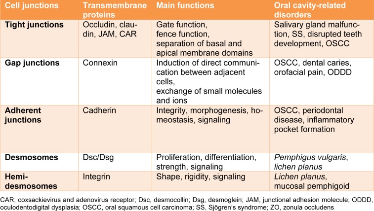
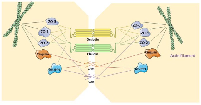
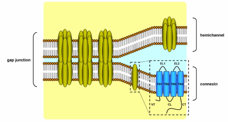
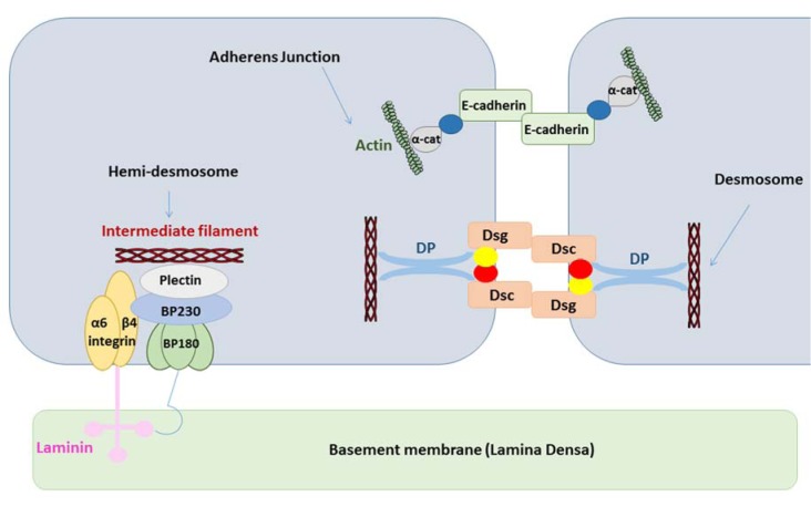
The oral cavity and its appendices are exposed to considerable environmental and mechanical stress. Cell junctions play a pivotal role in this context. Among those, gap junctions permit the exchange of compounds between cells, thereby controlling processes such as cell growth and differentiation. Tight junctions restrict paracellular transportation and inhibit movement of integral membrane proteins between the different plasma membrane poles. Adherens junctions attach cells one to another and provide a solid backbone for resisting to mechanistical stress. The integrity of oral mucosa, normal tooth development and saliva secretion depend on the proper function of all these types of cell junctions. Furthermore, deregulation of junctional proteins and/or mutations in their genes can alter tissue functioning and may result in various human disorders, including dental and periodontal problems, salivary gland malfunction, hereditary and infectious diseases as well as tumorigenesis. The present manuscript reviews the role of cell junctions in the (patho)physiology of the oral cavity and its appendices, including salivary glands.
Keywords: anchoring junction; gap junction; oral disease; oral health; tight junction.
Figures
References
-
- About I, Bottero M-J, de Denato P, Camps J, Franquin J-C, Mitsiadis TA. Human dentin production in vitro. Exp Cell Res. 2000;258:33–41. - PubMed
-
- About I, Proust J-P, Raffo S, Mitsiadis TA, Franquin J-C. In vivo and in vitro expression of connexin 43 in human teeth. Connec Tissue Res. 2002;43:232–237. - PubMed
-
- Ahmadian E, Eftekhari A, Samiei M, Maleki Dizaj S, Vinken M. The role and therapeutic potential of connexins, pannexins and their channels in Parkinson's disease. Cell Signal. 2019;58:111–118. - PubMed
-
- Amagai M, Matsuyoshi N, Wang ZH, Andl C, Stanley JR. Toxin in bullous impetigo and staphylococcal scalded-skin syndrome targets desmoglein 1. Nat Med. 2000;6:1275. - PubMed
-
- Anderson JM. Molecular structure of tight junctions and their role in epithelial transport. Physiology. 2001;16:126–130. - PubMed
Publication types
LinkOut - more resources
Full Text Sources
Miscellaneous