. 2019 May 29:10:622.
doi: 10.3389/fphys.2019.00622. eCollection 2019.

Secondary Placental Defects in Cxadr Mutant Mice

Affiliations

Secondary Placental Defects in Cxadr Mutant Mice

Jennifer E Outhwaite et al. Front Physiol. .

Abstract

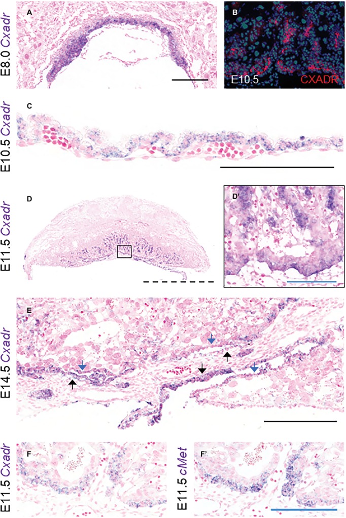
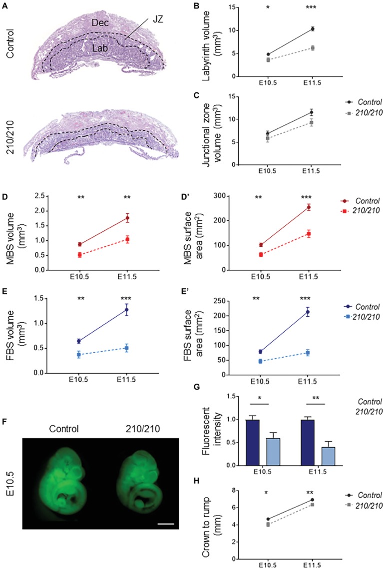
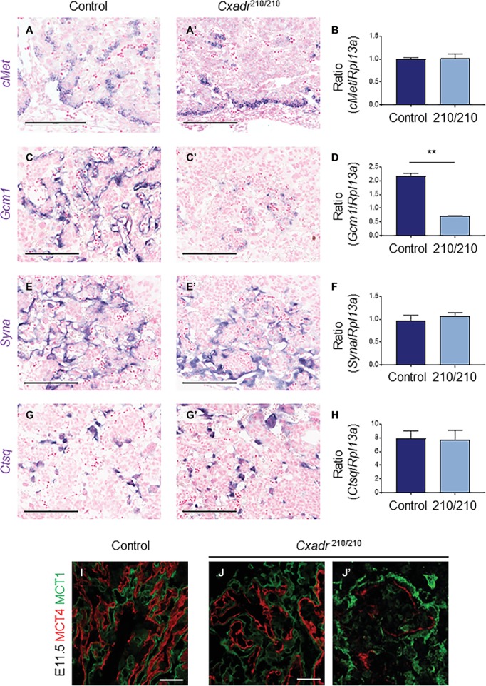
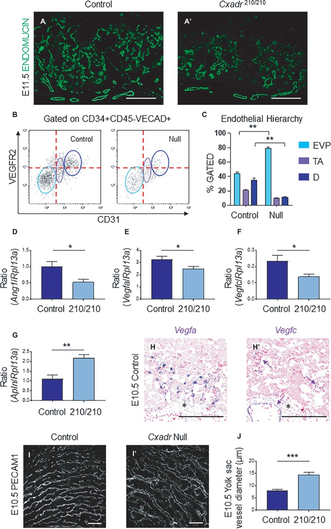
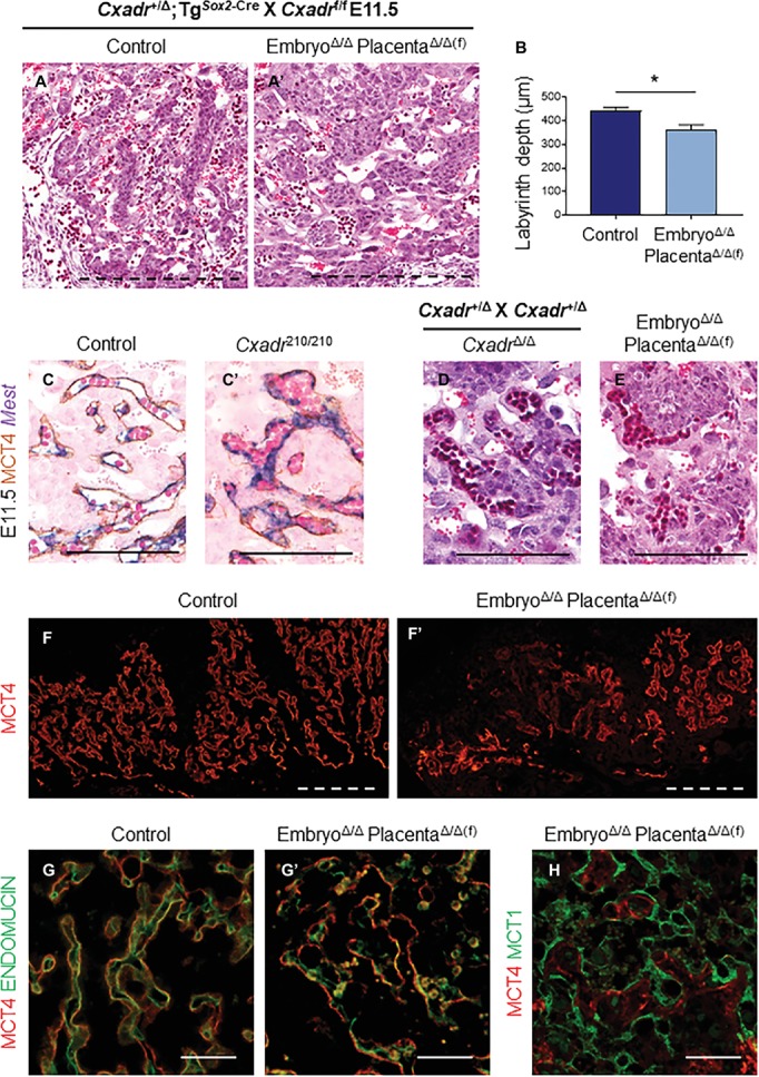
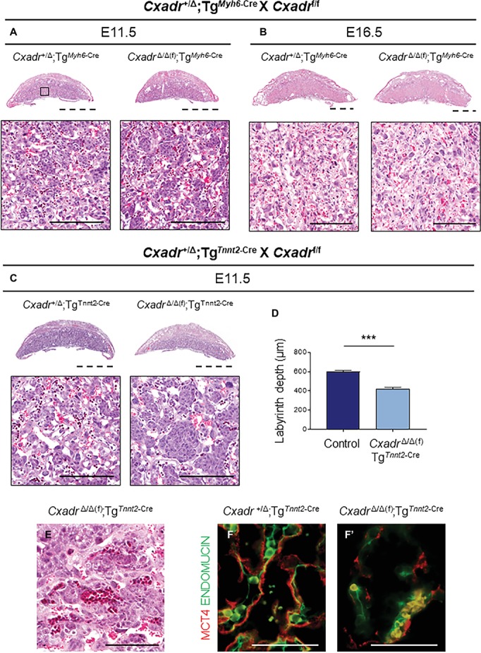
The Coxsackie virus and adenovirus receptor (CXADR) is an adhesion molecule known for its role in virus-cell interactions, epithelial integrity, and organogenesis. Loss of Cxadr causes numerous embryonic defects in mice, notably abnormal development of the cardiovascular system, and embryonic lethality. While CXADR expression has been reported in the placenta, the precise cellular localization and function within this tissue are unknown. Since impairments in placental development and function can cause secondary cardiovascular abnormalities, a phenomenon referred to as the placenta-heart axis, it is possible placental phenotypes in Cxadr mutant embryos may underlie the reported cardiovascular defects and embryonic lethality. In the current study, we determine the cellular localization of placental Cxadr expression and whether there are placental abnormalities in the absence of Cxadr. In the placenta, CXADR is expressed specifically by trophoblast labyrinth progenitors as well as cells of the visceral yolk sac (YS). In the absence of Cxadr, we observed altered expression of angiogenic factors coupled with poor expansion of trophoblast and fetal endothelial cell subpopulations, plus diminished placental transport. Unexpectedly, preserving endogenous trophoblast Cxadr expression revealed the placental defects to be secondary to primary embryonic and/or YS phenotypes. Moreover, further tissue-restricted deletions of Cxadr suggest that the secondary placental defects are likely influenced by embryonic lineages such as the fetal endothelium or those within the extraembryonic YS vascular plexus.

Keywords: Cxadr; fetal circulation; fetal heart; placenta; placenta-heart axis.


Figures

Figure 1
Figure 1
Cxadr is expressed in chorion, labyrinth trophoblast, and extraembryonic yolk sac. (A) In situ hybridization of Cxadr in basal chorion cells at E8.0. (B) Immunostaining for CXADR in placental labyrinth at E10.5. (C) Cxadr expression in visceral endoderm cells of the yolk sac at E10.5. (D) Expression of Cxadr in basal labyrinth cells and in clusters of trophoblast cells adjacent to fetal vessels at E11.5. (D’) Higher magnification image of inset in D. (E) Cxadr expression is evident in cells of the intraplacental yolk sac (IPYS) at E14.5. Parietal (blue arrow) and visceral (black arrow) IPYS layers are indicated. (F,F’) In situ hybridization of Cxadr (F) and cMet (F’) in basal chorion cells at E11.5. Scale bars: 100 μm (solid blue), 200 μm (solid black), and 2 mm (dashed).
Figure 2
Figure 2
CXADR is required for proper placental development. (A) Hematoxylin and eosin stained histological sections of Cxadr+/+ and Cxadr210/210 placentas at E11.5. Maternal decidua (Dec), labyrinth (Lab), and junctional zone (JZ) are indicated by dashed outlines. (B) Quantification of labyrinth volume. Plots represent the mean ± SEM for a minimum of n = 7 placentas per genotype. (C) Quantification of junctional zone volume. (D,D’) Quantification of maternal blood space volume (D) and surface area (D’). (E) Quantification of fetal blood space volume (E) and surface area (E’). (F,G) Whole-mount images of trans-placental passage of rhodamine 123 in embryos at E10.5 (F) and quantification (G) of fluorescent intensity. Plots represent the mean ± SEM for a minimum of n = 6 embryos per genotype. (H) Measure of crown to rump length. *p < 0.05, **p < 0.01, ***p < 0.001. Scale bar: 1 mm.
Figure 3
Figure 3
CXADR is required for appropriate expansion of labyrinth trophoblast cell types. In situ hybridization in Cxadr+/+ and Cxadr210/210 placentas at E11.5 for cMet (A,A’), Gcm1 (C,C’), Syna (E,E’), and Ctsq (G,G’). qRT-PCR analysis for cMet (B), Gcm1 (D), Syna (F), and Ctsq (H) relative to Rpl13a. Plots represent the mean ± SEM for a minimum of n = 7 placentas per genotype. (I,J) Immunofluorescence of MCT1 (green) marking syncytial layer I cells, and MCT4 (red) marking syncytial layer II cells in Cxadr+/+ (I) and Cxadr210/210 (J) labyrinth vessels at E11.5. (J’) Higher magnification image of Cxadr210/210 peripheral labyrinth vessel loss. **p < 0.01. Scale bars: 200 μm (black) and 50 μm (white).
Figure 4
Figure 4
Cxadr null mice display impaired vascular invasion and defective IHM morphogenesis. (A) Immunofluorescence of ENDOMUCIN (green) marking fetal endothelial cells in Cxadr+/+ (A) and Cxadr210/210 (A’) labyrinth sections at E11.5. (B) CD34+ CD45− VECAD+ gated labyrinth cells reveal distinct endothelial populations in Cxadr+/+ and CxadrΔ/Δ samples, based on VEGFR2 and CD31 expression. From left to right: endovascular progenitors (EVP), transit amplifying (TA), FIGURE 4and differentiated (D) endothelial cells. Minimum of n = 2 labyrinths per genotype were pooled per litter, and comparisons were made between n = 3 litters. (C) Endothelial hierarchy within each gated population/genotype. (D–F) qRT-PCR analysis for Ang1, Vegfa, and Vegfc expression in E11.5 placentas relative to Rpl13a. Plots represent the mean ± SEM for a minimum of n = 7 placentas per genotype. (G) qRT-PCR analysis for Apln expression in E11.5 placentas relative to Rpl13a. Plots represent the mean ± SEM for a minimum of n = 7 placentas per genotype. (H) In situ hybridization of Vegfa in labyrinth cells adjacent to fetal vessels at E10.5. (H’) In situ hybridization of Vegfc in some (arrow), not all, fetal endothelial cells of the labyrinth and allantois at E10.5. (I) Immunofluorescence of PECAM1 marking endothelial cells in Cxadr+/+ (I) and CxadrΔ/Δ (I’) flat mount yolk sacs at E10.5. (J) Quantification of yolk sac vessel diameter at E10.5. Plots represent the mean ± SEM for n = 5 yolk sacs per genotype. *p < 0.05, **p < 0.01, ***p < 0.001. Scale bars: 200 μm (white) and 300 μm (black).
Figure 5
Figure 5
Placental defects in Cxadr null mice are secondary to embryo development. (A) Hematoxylin and eosin stained histological sections of control (Cxadr+/Δ;TgSox2-Cre) (A) and CxadrΔ/Δ(f);TgSox2-Cre (A’) labyrinths at E11.5. (B) Average labyrinth depth. Plots represent the mean ± SEM for n = 4 placentas per genotype. (C) In situ hybridization for Mest and immunohistochemistry for MCT4 in Cxadr+/+ (C) and Cxadr210/210 (C’) peripheral interhaemal membrane structures at E11.5. (D,E) Hematoxylin and eosin stained histological sections of CxadrΔ/Δ (D) and CxadrΔ/Δ(f);TgSox2-Cre (E) peripheral labyrinth fetal vessels at E11.5. (F) Immunofluorescence of MCT4, marking syncytial layer II, in control (Cxadr+/Δ;TgSox2-Cre) (F) and mutant (CxadrΔ/Δ(f);TgSox2-Cre) (F’) E11.5 placentas. (G) High magnification images showing immunofluorescence of MCT4 (red) and ENDOMUCIN (green), marking fetal endothelial cells, in control (Cxadr+/Δ;TgSox2-Cre) (G) and mutant (CxadrΔ/Δ(f);TgSox2-Cre) (G’) labyrinth sections at E11.5. (H) Immunofluorescent detection of MCT4 (red) and MCT1 (green), marking syncytial layer I, in E11.5 CxadrΔ/Δ(f);TgSox2-Cre labyrinth sections. *p < 0.05. Scale bars: 300 μm (dashed black), 100 μm (solid black), 200 μm (dashed white), and 50 μm (solid white).
Figure 6
Figure 6
Heart specific deletion of Cxadr does not impact placental development. (A,B) Hematoxylin and eosin stained histological sections of control (Cxadr+/Δ;TgMyh6-Cre) and mutant (CxadrΔ/Δ(f);TgMyh6-Cre) placentas at E11.5 (A) and E16.5 (B). Higher magnification of inset depicts close up of labyrinth. (C) Hematoxylin and eosin stained histological sections of control (Cxadr+/Δ;TgTnnt2-Cre) and mutant (CxadrΔ/Δ(f);TgTnnt2-Cre) placentas at E11.5. (D) E11.5 control (Cxadr+/Δ;TgTnnt2-Cre) and mutant (CxadrΔ/Δ(f);TgTnnt2-Cre) labyrinth depths. Plots represent the mean ± SEM for n = 4 placentas per genotype. (E) Hematoxylin and eosin stained histological section of CxadrΔ/Δ(f); TgSox2-Cre peripheral labyrinth fetal vessels at E11.5. (F) High magnification images showing immunofluorescence of MCT4 (red), marking syncytial layer II, and ENDOMUCIN (green), marking fetal endothelial cells, in control (Cxadr+/Δ;TgTnnt2-Cre) (F) and mutant (CxadrΔ/Δ(f);TgTnnt2-Cre) (F’) labyrinth sections at E11.5. ***p < 0.001. Scale bars: 2 mm (dashed black), 100 μm (solid black), and 50 μm (solid white).
Figure 7
Figure 7
Timeline of placental, yolk sac (YS), and cardiac developmental milestones occurring between E7.5 and E11.5.

Similar articles

Cited by

References

    1. Adams R. H., Porras A., Alonso G., Jones M., Vintersten K., Panelli S., et al. . (2000). Essential role of p38α MAP kinase in placental but not embryonic cardiovascular development. Mol. Cell 6, 109–116. 10.1016/S1097-2765(05)00014-6, PMID: - DOI - PubMed
    1. Ahmed A., Perkins J. (2000). Angiogenesis and intrauterine growth restriction. Best Pract. Res. Clin. Obstet. Gynaecol. 14, 981–998. 10.1053/beog.2000.0139, PMID: - DOI - PubMed
    1. Asher D. R., Cerny A. M., Weiler S. R., Horner J. W., Keeler M. L., Neptune M. A., et al. . (2005). Coxsackievirus and adenovirus receptor is essential for cardiomyocyte development. Genesis 42, 77–85. 10.1002/gene.20127, PMID: - DOI - PubMed
    1. Barak Y., Nelson M. C., Ong E. S., Jones Y. Z., Ruiz-Lozano P., Chien K. R., et al. . (1999). PPAR gamma is required for placental, cardiac, and adipose tissue development. Mol. Cell 4, 585–595. 10.1016/S1097-2765(00)80209-9, PMID: - DOI - PubMed
    1. Bergelson J. M., Cunningham J. A., Droguett G., Kurt-Jones E. A., Krithivas A., Hong J. S., et al. . (1997). Isolation of a common receptor for coxsackie B viruses and adenoviruses 2 and 5. Science 275, 1320–1323. 10.1126/science.275.5304.1320, PMID: - DOI - PubMed

LinkOut - more resources