. 2019 Jul 9:10:1499.
doi: 10.3389/fmicb.2019.01499. eCollection 2019.

The Response of Cupriavidus metallidurans CH34 to Cadmium Involves Inhibition of the Initiation of Biofilm Formation, Decrease in Intracellular c-di-GMP Levels, and a Novel Metal Regulated Phosphodiesterase

Affiliations

The Response of Cupriavidus metallidurans CH34 to Cadmium Involves Inhibition of the Initiation of Biofilm Formation, Decrease in Intracellular c-di-GMP Levels, and a Novel Metal Regulated Phosphodiesterase

Pablo Alviz-Gazitua et al. Front Microbiol. .

Erratum in

Abstract

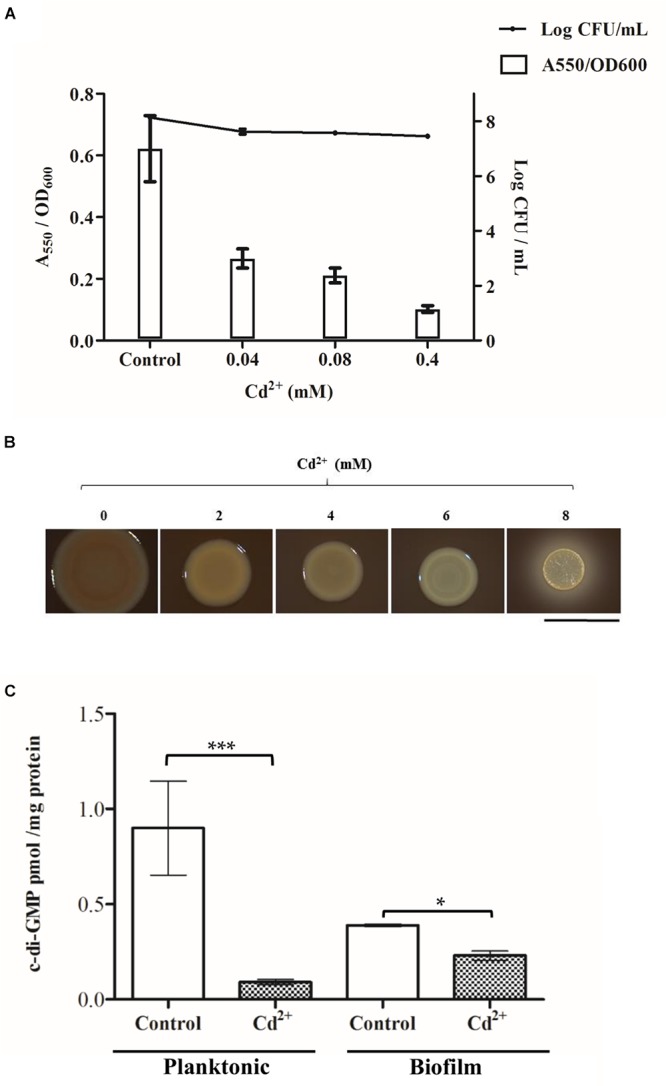
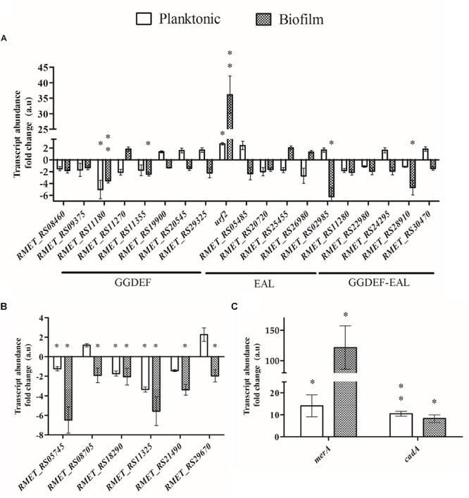
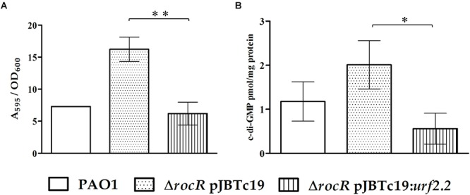
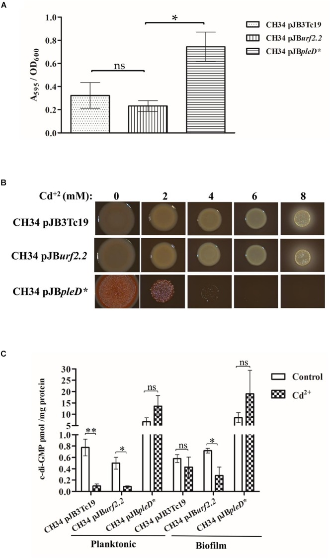
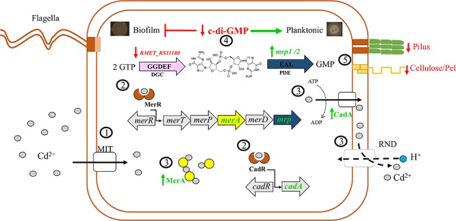
Cadmium is a highly toxic heavy metal for biological systems. Cupriavidus metallidurans CH34 is a model strain to study heavy metal resistance and bioremediation as it is able to deal with high heavy metal concentrations. Biofilm formation by bacteria is mediated by the second messenger bis-(3'-5')-cyclic dimeric guanosine monophosphate (c-di-GMP). The aim of this study was to characterize the response of C. metallidurans CH34 planktonic and biofilm cells to cadmium including their c-di-GMP regulatory pathway. Inhibition of the initiation of biofilm formation and EPS production by C. metallidurans CH34 correlates with increased concentration of cadmium. Planktonic and biofilm cells showed similar tolerance to cadmium. During exposure to cadmium an acute decrease of c-di-GMP levels in planktonic and biofilm cells was observed. Transcription analysis by RT-qPCR showed that cadmium exposure to planktonic and biofilm cells induced the expression of the urf2 gene and the mercuric reductase encoding merA gene, which belong to the Tn501/Tn21 mer operon. After exposure to cadmium, the cadA gene involved in cadmium resistance was equally upregulated in both lifestyles. Bioinformatic analysis and complementation assays indicated that the protein encoded by the urf2 gene is a functional phosphodiesterase (PDE) involved in the c-di-GMP metabolism. We propose to rename the urf2 gene as mrp gene for metal regulated PDE. An increase of the second messenger c-di-GMP content by the heterologous expression of the constitutively active diguanylate cyclase PleD correlated with an increase in biofilm formation and cadmium susceptibility. These results indicate that the response to cadmium in C. metallidurans CH34 inhibits the initiation of biofilm lifestyle and involves a decrease in c-di-GMP levels and a novel metal regulated PDE.

Keywords: Cupriavidus metallidurans; PleD; biofilm; c-di-GMP; cadmium; mer gene; phosphodiesterase; urf2 gene.


Figures

Figure 1
Figure 1
Effect of cadmium on C. metallidurans CH34 biofilm formation and c-di-GMP levels. (A) Cell attachment and cell viability after 15 h of incubation in LPTMS broth at 30°C. The means of three independent experiments and standard deviations are shown. (B) Macrocolonies grown on Congo Red LB agar plates at different cadmium concentrations. Images were taken after 5 days of incubation at room temperature. The red staining intensity of the macrocolony indicates the presence of EPS. The bar represents 1 cm. (C) c-di-GMP levels after 2 mM cadmium exposure during 45 min in LPTMS broth (planktonic) or on agar (biofilm). The c-di-GMP concentration was normalized with respect to total protein content in each sample. The means of three independent experiments and standard deviations are shown. Significant differences assessed by t-test: P < 0.05, ∗∗P < 0.01, ∗∗∗P < 0.001.
Figure 2
Figure 2
Domain-level depiction of c-di-GMP metabolism proteins from C. metallidurans CH34. Proteins with GGDEF/EAL domains that are catalytically active according to bioinformatic analyses are shown. (A) Diguanylate cyclases. (B) Phosphodiesterases. (C) Hybrid GGDEF/EAL proteins.
Figure 3
Figure 3
Effect of cadmium on the transcription of c-di-GMP metabolic and heavy metal resistance genes in planktonic and biofilm C. metallidurans CH34 cells. The transcription was measured after exposure to cadmium (2 mM) for 45 min. (A) Genes involved in c-di-GMP metabolism. The term urf2 represents both urf2.1 and urf2.2 genes. (B) Genes encoding c-di-GMP effectors. (C) Mercury and cadmium resistance genes. Each bar represents the mean of values of three independent experiments, each analyzed in duplicate, and standard deviations. Significant differences in expression to the control condition in absence of cadmium, assessed by t-test, are shown in asterisk codes: P < 0.05, ∗∗P < 0.01.
Figure 4
Figure 4
Complementation effects in P. aeruginosa PAO1 ΔrocR with pJBurf2.2. (A) Biofilm formation at 6 h of incubation in 96-well plates without cadmium. (B) The c-di-GMP content in planktonic cells at 6 h of incubation in Erlenmeyer flasks. The means of values of three independent experiments and standard deviations are shown. Significant differences assessed by t-test: P < 0.05, ∗∗P < 0.01.
Figure 5
Figure 5
Effect of urf2.2 overexpression and pleD heterologous expression on C. metallidurans CH34 biofilm formation and c-di-GMP metabolism. (A) Biofilm formation in the absence of cadmium. The means of three independent experiments and standard deviations are shown. Significant differences assessed by t-test: P < 0.05; ns, not significant. (B) Macrocolonies morphotype in the presence of cadmium. The red staining of the macrocolony indicates its EPS content. (C) Response in c-di-GMP levels after cadmium exposure. The means of three independent experiments and standard deviations are shown. Significant differences assessed by t-test: P < 0.05, ∗∗P < 0.01; ns, non-significant.
Figure 6
Figure 6
Proposed model of the C. metallidurans CH34 and its c-di-GMP signaling pathway in response to cadmium. 1, Cadmium ions (gray circles) enter the cell through metal ion transporters (MIT). 2, Cadmium induces gene expression of detoxification systems CadA, MerA, and RND antiporter pumps. Cadmium ions upregulate the phosphodiesterases (PDEs) (mrp1 and mrp2 genes) encoded in the mercury resistance operons Tn4378 and Tn4380 and downregulate the diguanylate cyclase (RMET_RS11180). 3, MerA proteins (yellow circles) may act as chelator proteins of free cadmium cooperating with canonical detoxification systems. Efflux of cadmium is facilitated by CadA and RND antiporter pumps. 4, Upregulation of Mrp1/2 and downregulation of RMET_RS11180 protein decreased c-di-GMP levels. 5, The drop of c-di-GMP levels inhibits the cell adhesion (pilus) and extracellular polysaccharide synthesis (cellulose and Pel) that may favor the planktonic lifestyle. Vertical arrows stand for upregulation (green) and downregulation (red) of molecules.

References

    1. Agulló L., Romero-Silva M. J., Domenech M., Seeger M. (2017). p-Cymene promotes its catabolism through the p-cymene and the p-cumate pathways, activates a stress response and reduces the biofilm formation in Burkholderia xenovorans LB400. PLoS One 12:e0169544. 10.1371/journal.pone.0169544 - DOI - PMC - PubMed
    1. Altschul S. F., Madden T. L., Schäffer A. A., Zhang J., Zhang Z., Miller W., et al. (1997). Gapped BLAST and PSI-BLAST:a new generation of protein database search programs. Nucleic Acids Res. 25 3389–3402. 10.1093/nar/25.17.3389 - DOI - PMC - PubMed
    1. Begg S. L., Eijkelkamp B. A., Luo Z., Couñago R. M., Morey J. R., Maher M. J., et al. (2015). Dysregulation of transition metal ion homeostasis is the molecular basis for cadmium toxicity in Streptococcus pneumoniae. Nat. Commun. 6:6418. 10.1038/ncomms7418 - DOI - PMC - PubMed
    1. Bernstein J. A., Khodursky A. B., Lin P.-H., Lin-Chao S., Cohen S. N. (2002). Global analysis of mRNA decay and abundance in Escherichia coli at single-gene resolution using two-color fluorescent DNA microarrays. Proc. Natl. Acad. Sci. U.S.A. 99 9697–9702. 10.1073/pnas.112318199 - DOI - PMC - PubMed
    1. Biemans-Oldehinkel E., Mahmood N. A. B. N., Poolman B. (2006). A sensor for intracellular ionic strength. Proc. Natl. Acad. Sci. U.S.A. 103 10624–10629. 10.1073/pnas.0603871103 - DOI - PMC - PubMed