

Delayed evolving epidural hematoma in the setting of a depressed skull fracture: A case report and review of the literature
- PMID: 31338404
- PMCID: PMC6606925
- DOI: 10.1016/j.tcr.2019.100193
Delayed evolving epidural hematoma in the setting of a depressed skull fracture: A case report and review of the literature
Abstract
Background: Historically, in the pediatric population, there is a highly selective approach for repeat imaging given the risk of radiation and costs. In the lieu of this, frequent neurological checks and even ICP monitoring has been used as an adjunct, although not always successful. We present a case of a pediatric patient with a late evolving epidural hematoma in the setting of a depressed skull fracture, and present an argument for serial CT imaging in a select patient population similar to his.
Objective: Discuss the unique presentation, diagnosis, and management of an expanding epidural hematoma in a pediatric patient with a depressed skull fracture and the need for aggressive repeat imaging in this setting.
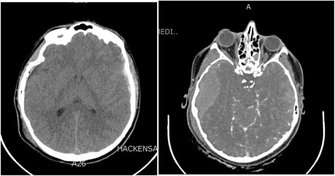
Case report: Patient is a 15-year-old boy who presented to our trauma bay after being the victim of a hit and run while skateboarding. His injuries included a depressed comminuted skull fracture and bilateral SDH. Additionally, a stat CT angiogram was obtained due to a basilar skull fracture. A rapidly evolving EDH with impending herniation was found, which was nearly fatal and was not present on the initial CT scan. He required emergent evacuation with a hemi-craniectomy where he was found to have a laceration of his dural vessels as well as his middle meningeal artery. Post operatively he did well and regained full neurologic function.
Conclusion: We presented a case of a pediatric patient with a late evolving epidural hematoma seen on repeat CT imaging. In the setting of a depressed skull fracture, hemorrhage from this source is likely to be missed on initial CT imaging. Frequent neurochecks or ICP monitoring may not be possible in this population encouraging the need for more aggressive repeat imaging.
Figures
References
-
- Hill E.P., Stiles P., Reyes J., Nold R.J., Helmer S.D., Haan J.M. Repeat head imaging in blunt pediatric trauma patients. Is it necessary? J. Trauma Acute Care Surg. 2017;82(5):896–900. - PubMed
-
- Stanley R.M., Johnson M., Vanc C., Bajaj L., Babcock L. Challenges enrolling children into traumatic brain injury trials: an observational study. Acad. Emerg. Med. 2016;24(1):31–39. - PubMed
-
- Tunik M.G., Powell E.C., Mahajan P., Schunk J.E., Jacobs E. Clinical presentations and outcomes of children with basilar skull fractures after blunt head trauma. Ann. Emerg. Med. 2016;68(4):431–440. - PubMed
-
- Blackwood B., Bean J., Sadecki-Lund C., Helenowski I., Kabre R., Hunter C. Observation for isolated traumatic skull fractures in the pediatric population: unnecessary and costly. J. Pediatr. Surg. 2016;51(4):654–658. - PubMed
Publication types
LinkOut - more resources
Full Text Sources
