Review
. 2019 Jul 24;10(1):67.
doi: 10.1186/s13244-019-0752-4.

MRI of the axial skeleton in spondyloarthritis: the many faces of new bone formation

Affiliations
Review

MRI of the axial skeleton in spondyloarthritis: the many faces of new bone formation

Frederiek Laloo et al. Insights Imaging. .

Abstract

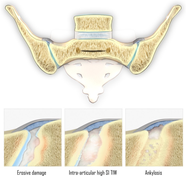
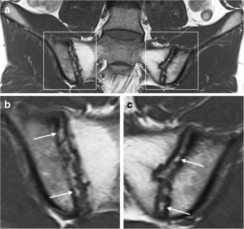
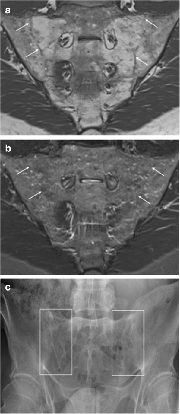
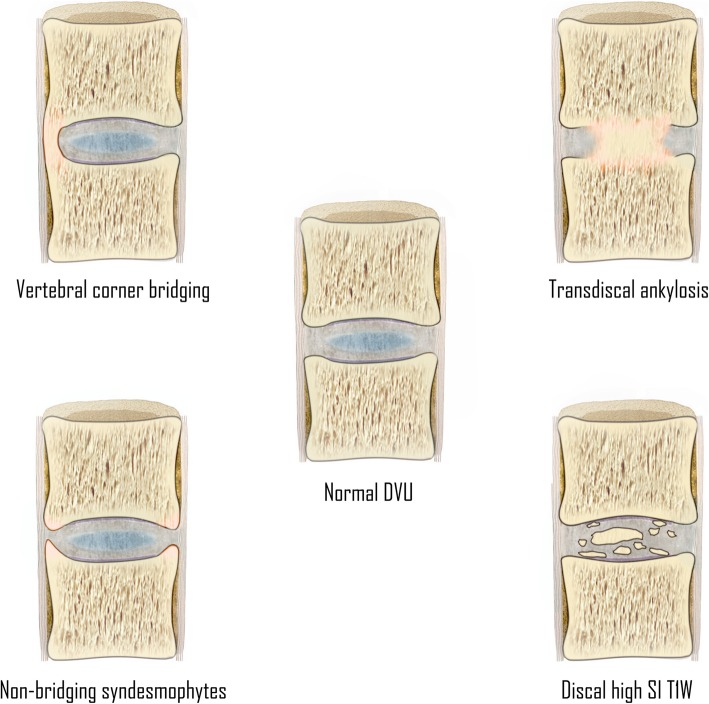
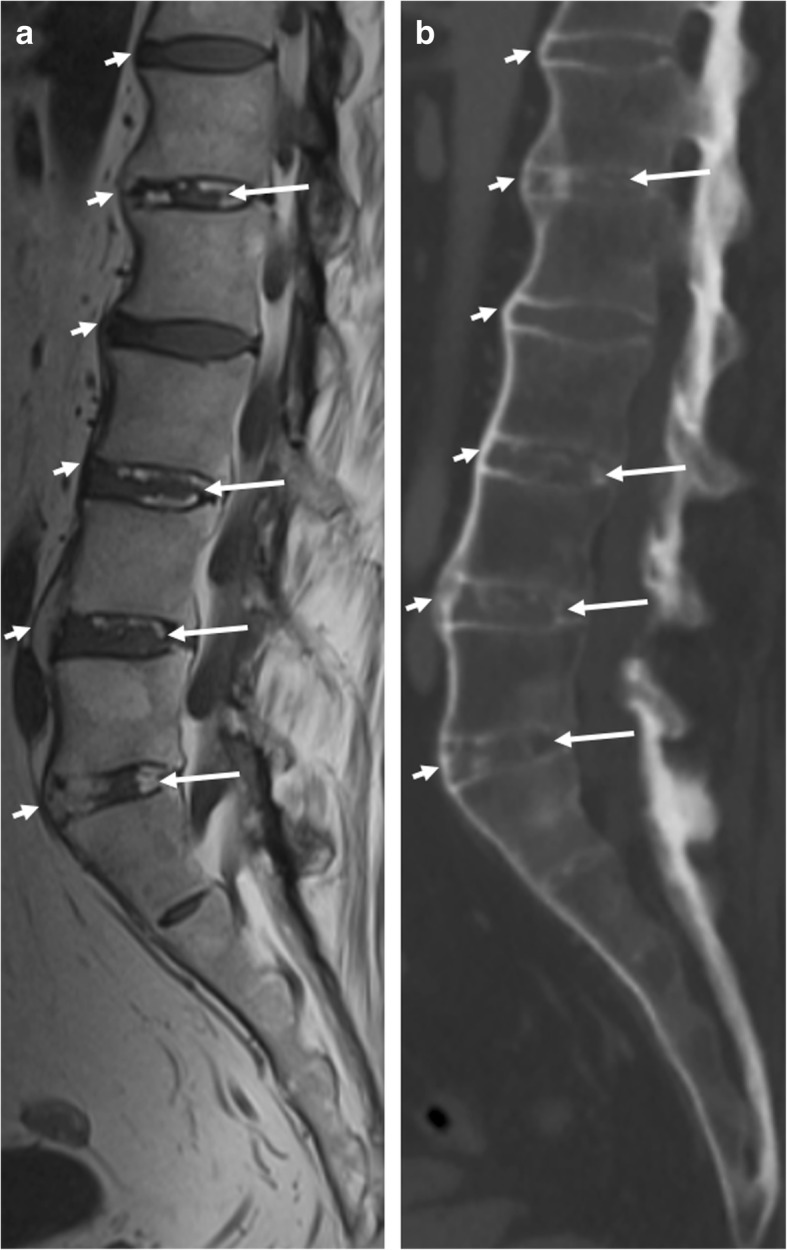
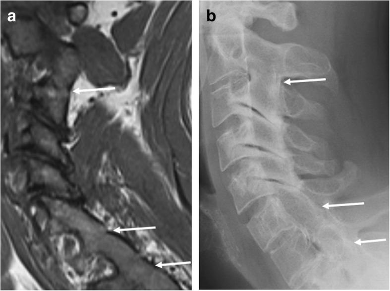
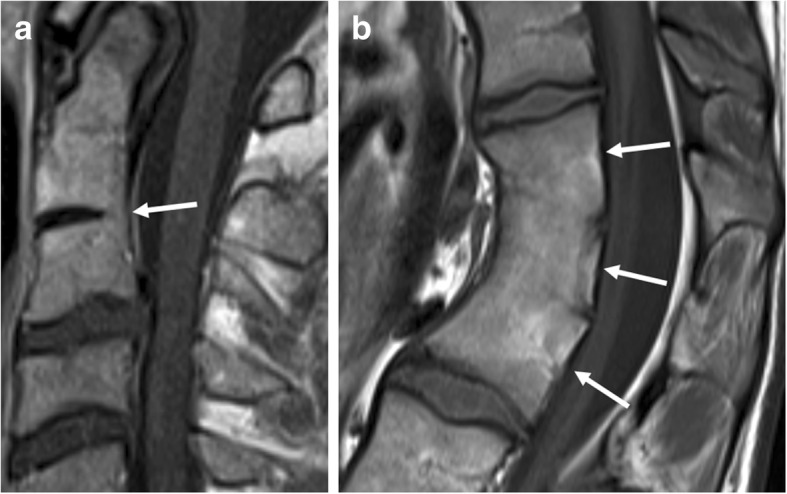
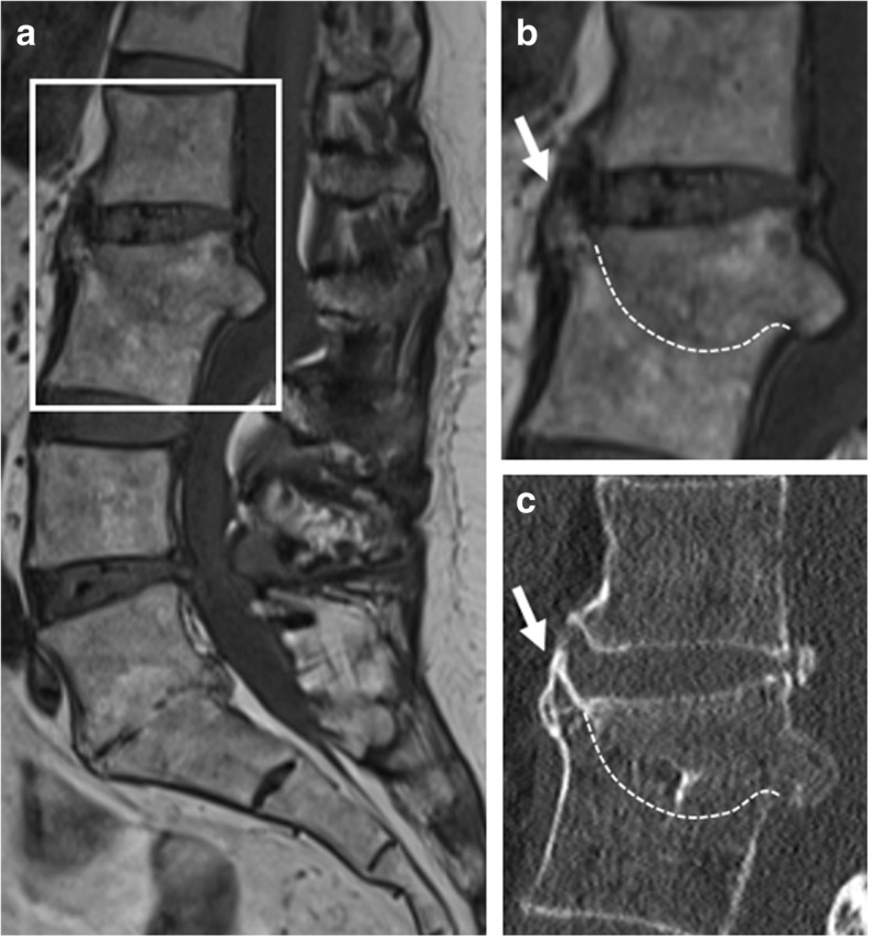
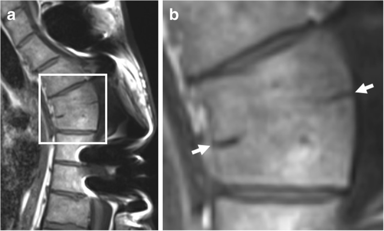
Spondyloarthritis has two hallmark features: active inflammation and structural lesions with new bone formation. MRI is well suited to assess active inflammation, but there is increasing interest in the role of structural lesions at MRI. Recent MRI studies have examined the established features of new bone formation and demonstrated some novel features which show diagnostic value and might even have potential as possible markers of disease progression. Although MRI is not the first imaging modality that comes into mind for assessment of bony changes, these features of new bone formation can be detected on MRI-if one knows how to recognize them. This review illustrates the MRI features of new bone formation and addresses possible pitfalls.

Keywords: Ankylosis; Magnetic resonance imaging; Sacroiliac joint; Spine; Spondyloarthritis.


Conflict of interest statement

The authors declare that they have no competing interests.

Figures

Fig. 1
Fig. 1
Features associated with new bone formation in the sacroiliac joints of patients with SpA. Note: erosive damage occurs before new bone formation. (SI T1W = signal intensity on T1-weighted MR images)
Fig. 2
Fig. 2
Intra-articular high signal intensity in the sacroiliac joints space on T1-weighted MR images. Coronal oblique T1-weighted MR images acquired in a 25-year-old man with SpA show high intra-articular signal intensity (arrows). This high signal intensity is clearly filling up the eroded iliac bone on the right (i.e., “backfill”). Extensive fatty degeneration of the bone marrow of the sacral side of the sacroiliac joints is present, i.e., post-inflammatory structural changes. Note: b and c are enlargements of the regions outlined in a
Fig. 3
Fig. 3
Ankylosis of the sacroiliac joints. Coronal oblique MR images acquired in a 53-year-old man with SpA show bony fusion of the sacroiliac joints as (a) high signal intensity on T1-weighted imaging (fatty degeneration) and (b) low signal intensity on STIR imaging. A sclerotic remnant of the sacroiliac joints is highlighted (arrows). c Radiography of the sacroiliac joints confirms these structural bony changes
Fig. 4
Fig. 4
Features associated with new bone formation at the disco-vertebral unit of patients with SpA. (DVU = disco-vertebral unit; SI T1W = signal intensity on T1-weighted MR images)
Fig. 5
Fig. 5
Discal high signal intensity on T1-weighted MR images. a Sagittal T1-weighted MR images of the lumbosacral spine acquired in a 45-year-old man with SpA show signal intensity (long arrows) similar to that of adipose tissue within the intervertebral disc. b Computed tomography (CT) of the same intervertebral discs show discal calcification (long arrows). Also, notice the discordance in the visibility between CT and MRI for the evaluation of anterior bridging syndesmophytes (short arrows)
Fig. 6
Fig. 6
Non-bridging syndesmophytes. Sagittal T1-weighted MR image shows anterior syndesmophytes (arrows) in a patient without SpA. No other disco-vertebral units besides Th3-Th4 and Th4-Th5 show bony changes. Note: b is an enlargement of the region outlined in a
Fig. 7
Fig. 7
Vertebral corner bridging. a Sagittal T1-weighted MR imaging acquired in a 57-year-old man with SpA shows bridging syndesmophytes (arrows), and (b) radiography of the same vertebrae confirms these vertebral corner bridges (arrows) resulting in a “bamboo spine” configuration. Note that the smaller anterior syndesmophytes are more clearly visualized on radiograph than MRI, which is typical
Fig. 8
Fig. 8
Extensive—and several types of—new bone formation in one patient with SpA. Sagittal T1-weighted MR image shows how bony fusion of the manubriosternal joint can be observed on sagittal imaging of the spine in a routine SpA protocol (rectangle). Also, notice the other types of new bone formation: syndesmophytes (dotted arrow), discal high signal intensity and/ or progressive trandiscal ankylosis (short arrow), and osteophytes (long arrow). This image demonstrates that osteophytes can be seen in patients with SpA at levels otherwise unaffected by SpA, perhaps due to increased mechanical loads on the remaining functional disco-vertebral units
Fig. 9
Fig. 9
Ankylosis of the facet joints. a Sagittal T1-weighted MR image acquired in a 34-year-old man with SpA shows ankylosis of the facet joints of C2-C3, C6-C7, and C7-Th1 (arrows). b Radiography of the cervical spine confirms this bony fusion of these joints (arrows)
Fig. 10
Fig. 10
Pitfalls in imaging and diagnosis of new bone formation: DISH. a Sagittal T1-weighted MR image and (b) computed tomography (CT) show hyperproliferative ossification of the anterior longitudinal ligament, resulting in bulky horizontally oriented osteophytes (arrows) with—in this case—an average growth angle of > 45° from vertical. c, d Semicoronal T1-weighted MR images show bridging ossification (arrows) at the anterior and superior aspect of the sacroiliac joints
Fig. 11
Fig. 11
Pitfalls in imaging and diagnosis of new bone formation: congenital block vertebrae. Sagittal T1-weighted MR images show (a) partial congenital block vertebra of C2-C3 (arrow) and (b) complete congenital block vertebra consisting out of three vertebrae (arrows). Note the narrowed antero-posterior diameter in b, a typical sign of complete congenital fusion. Also, note that the height in b is less than the expected sum of three vertebrae and two intervertebral discs
Fig. 12
Fig. 12
Pitfalls in imaging and diagnosis of new bone formation: sequelae of infectious spondylodiscitis. a, b Sagittal T1-weighted MR images and (c) computed tomography (CT) show vertebral fusion (dotted line) of L2-L3 after destruction of the vertebrae and intervertebral disc due to tuberculous spondylodiscitis. Note that the protrusion of the remainder of the anterior corner of the L3 mimics plump syndesmophyte formation (short arrow). L5-S1 also shows signs of vertebral fusion after destruction of the intervertebral disc, mimicking transdiscal ankylosis as seen in SpA
Fig. 13
Fig. 13
Pitfalls in imaging and diagnosis of new bone formation: post-traumatic vertebral fusion. Sagittal T1-weighted MR images show interbody vertebral fusion years after severe spinal trauma, with discrete remnants of the intervertebral disc (arrows). This image suggests that the original injury included both vertebral endplates and the intervertebral disc. Also note that the height is less than the sum of the two vertebral bodies and the intervertebral disc, and antero-posterior diameter is not narrowed, two features which help differentiate this image from a congenital block vertebra. Note: b is an enlargement of the region outlined in a

References

    1. Braun J, Sieper J. Ankylosing spondylitis. Lancet. 2007;369:1379–1390. doi: 10.1016/S0140-6736(07)60635-7. - DOI - PubMed
    1. Ward MM, Deodhar A, Akl EA, et al. American College Of Rheumatology/Spondylitis Association of America/Spondyloarthritis research and treatment network 2015 recommendations for the treatment of ankylosing spondylitis and non-radiographic axial spondyloarthritis. Arthritis Rheumatol. 2016;68:282–298. doi: 10.1002/art.39298. - DOI - PMC - PubMed
    1. Lacout A, Rousselin B, Pelage J. CT and MRI of spine and sacroiliac involvement in spondylarthropathy. AJR Am J Roentgenol. 2008;191:1016–1023. doi: 10.2214/AJR.07.3446. - DOI - PubMed
    1. Dougados M, Baeten D. Spondyloarthritis. Lancet. 2011;377:2127–2137. doi: 10.1016/S0140-6736(11)60071-8. - DOI - PubMed
    1. Jans L, Van Langenhove C, Van Praet L, et al. Diagnostic value of pelvic enthesitis on MRI of the sacroiliac joints in spondyloarthritis. Eur Radiol. 2014;24:866–871. doi: 10.1007/s00330-013-3074-9. - DOI - PubMed

LinkOut - more resources