Review
. 2019 Sep;9(3):449-468.
doi: 10.1007/s13555-019-0309-y. Epub 2019 Jul 23.

Atrophic Dermatofibroma: A Comprehensive Literature Review

Affiliations
Review

Atrophic Dermatofibroma: A Comprehensive Literature Review

Philip R Cohen et al. Dermatol Ther (Heidelb). 2019 Sep.

Abstract

Introduction: An atrophic dermatofibroma is a benign fibrohistiocytic neoplasm. It typically presents as an asymptomatic patch with a depressed central area.

Methods: The PubMed database was used to search the following words: atrophic, dermatofibroma, elastic and fibers. The relevant papers and their references generated by the search were reviewed. Images of the clinical and pathological features of two patients with an atrophic dermatofibroma are presented. In addition, a comprehensive review of the characteristics of this unique dermatofibroma is provided.

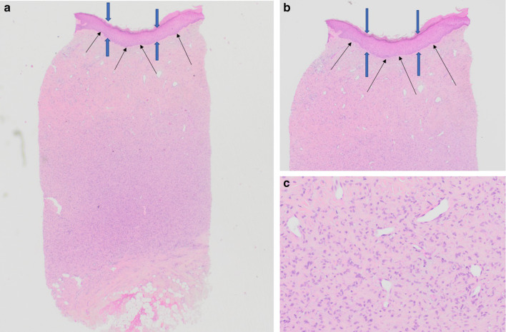
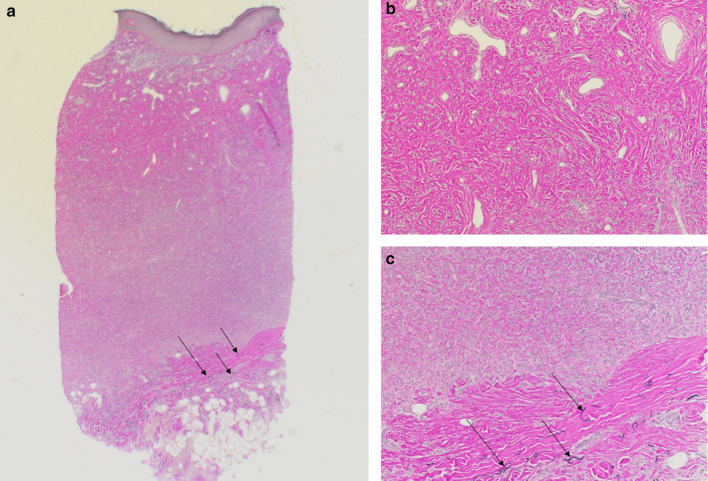
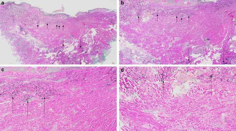
Results: An atrophic dermatofibroma has been reported in 102 patients: 53 women, 11 men and 38 individuals whose gender was not provided. It typically appeared as an asymptomatic solitary patch with a central umbilication-most commonly on the shoulder or lower extremity or back-of women aged 48 years or older. Dermoscopy typically showed white scar-like patches; a patchy pigment network was also noted in some lesions. The pathology of an atrophic dermatofibroma has the same features that can be observed in a common fibrous dermatofibroma; there is acanthosis, basal layer hyperpigmentation, and induction of basal cell carcinoma-like features, hair follicle formation or sebaceous hyperplasia in the epidermis and a proliferation of spindle-shaped fibroblasts in the dermis. However, atrophic dermatofibromas also demonstrate depression of the central surface and thinning of the dermis; in many cases, the dermal atrophy is at least 50%. Elastic fibers are either decreased or absent. Similar to non-atrophic dermatofibromas, the immunoperoxidase profile of atrophic dermatofibromas is factor XIIIa-positive and cluster of differentiation 34 (CD34)-negative. The pathogenesis of atrophic dermatofibromas remains to be established.

Conclusion: An atrophic dermatofibroma is an uncommon benign variant of a dermatofibroma. The diagnosis can be suspected based on clinical features and dermatoscopic findings. A biopsy of the lesion will confirm the diagnosis. Periodic evaluation of the lesion site is a reasonable approach to the management of the residual tumor.

Keywords: Atrophic; Depression; Dermatofibroma; Dermoscopy; Elastic; Fibers; Fibroblast; Men; Umbilication; Women.


Conflict of interest statement

Philip R. Cohen, Christof P. Erickson and Antoanella Calame have nothing to disclose with regards to the publication of this article. Philip R. Cohen is a member of the journal’s Editorial Board

Figures

Fig. 1
Fig. 1
Atrophic dermatofibroma: left shoulder of a 45-year-old Caucasian man. Distant (a) and closer (b) views of an asymptomatic lesion on the left shoulder of more than 3 years of duration. There was no prior history of trauma or injection to the site. As a youth, he had cystic acne. The lesion appeared as a 15 × 15-mm flesh-colored indurated patch surrounding a 10 × 10-mm telangiectatic depressed central area; the lesion is outlined by the purple lines (b). Squeezing the edges of the lesion between the examiner’s thumb and index finger produced a dimpling of the central portion of the lesion. The submitted clinical differential diagnosis was an atrophic dermatofibroma
Fig. 2
Fig. 2
Atrophic dermatofibroma on the left shoulder of a 45-year-old Caucasian man: pathology features of hematoxylin and eosin-stained sections. Low magnification (a) and higher magnification (b, c) views of a 3-mm punch biopsy from the central portion of the depressed area show epidermal acanthosis (between blue arrows) with basal layer hyperpigmentation (black arrows) (a, b). There is atrophy of the dermis, and the dermal tumor shows an increased number of fibroblasts with trapped collagen bundles in the periphery (hematoxylin and eosin: a ×2; b ×4; c ×20)
Fig. 3
Fig. 3
Atrophic dermatofibroma on the left shoulder of a 45-year-old Caucasian man: pathology features of Verhoeff-Van Gieson-stained sections. Low magnification (a) and higher magnification (b, c) views of a 3-mm punch biopsy from the central portion of the depressed area show an absence of elastic fibers in the tumor. However, elastic fibers (which stain black and are demonstrated by black arrows) can be noted in the deep dermis beneath the dermatofibroma (a, c) (Verhoeff-Van Gieson: a ×2; b ×20; c ×20)
Fig. 4
Fig. 4
Atrophic dermatofibroma: left upper back of a 64-year-old Caucasian man. Distant (a) and closer (b) views of an asymptomatic lesion on the left upper back of 1-year duration that had been noticed by his wife. There was no prior history of trauma or injection to the site. The lesion appeared as a 6 × 6-mm flesh-colored depressed area; the lesion is outlined by the purple lines (b). The submitted clinical differential diagnosis was an atrophic dermatofibroma
Fig. 5
Fig. 5
Atrophic dermatofibroma on the left upper back of a 64-year-old Caucasian man: pathology features of hematoxylin and eosin-stained sections. Low magnification (a) and higher magnification (bd) views of an 8-mm punch biopsy show a depressed area in the central portion of the specimen (between white arrows). The epidermis has seborrheic keratosis-like hyperplasia (between blue arrows) (a, b) and hyperpigmentation of the basal layer (black arrows) (ac). The center depression reveals atrophy of the dermis (a, b). The dermal tumor consists an increased number of fibroblasts with trapped collagen bundles in the periphery (ad) (Hematoxylin and eosin: a ×2; b ×4; c ×20; d ×10)
Fig. 6
Fig. 6
Atrophic dermatofibroma on the left upper back of a 64-year-old Caucasian man: pathology features of Verhoeff-Van Gieson-stained sections. Low magnification (a) and higher magnification (bd) views of an 8-mm punch biopsy show an absence of elastic fibers in more than 90% of the dermatofibroma. Elastic fibers (which stain black and are demonstrated by black arrows) are only present in the superficial portion of the tumor in the papillary dermis (ad). In addition, black staining of elastic fibers (black arrows) can be observed in the deep dermis beneath the dermatofibroma (a, b) (Verhoeff-Van Gieson: a ×2; b ×4; c ×10; d ×20)

References

    1. Canelas MM, Cardoso JC, Andrade PF, Reis JP, Tellechea O. Fibrous histiocytomas: histopathologic review of 95 cases. An Bras Dermatol. 2010;85:211–215. doi: 10.1590/S0365-05962010000200011. - DOI - PubMed
    1. Luzar B, Calonje E. Cutaneous fibrohistiocytic tumours—an update. Histopathology. 2010;56:148–165. doi: 10.1111/j.1365-2559.2009.03447.x. - DOI - PubMed
    1. Han TY, Chang HS, Lee JH, Lee WM, Son SJ. A clinical and histopathological study of 122 cases of dermatofibroma (benign fibrous histiocytoma) Ann Dermatol. 2011;23:185–192. doi: 10.5021/ad.2011.23.2.185. - DOI - PMC - PubMed
    1. Myers DJ, Fillman EP. Dermatofibroma. StatPearls. Treasure Island: StatPearls Publishing; 2018. p. 27.
    1. Beatrous SV, Riahi RR, Grisoli SB, Cohen PR. Associated conditions in patients with multiple dermatofibromas: case reports and literature review. Dermatol Online J. 2017;23(9):13030. - PubMed

LinkOut - more resources