Review
. 2019 Aug;20(8):1247-1265.
doi: 10.3348/kjr.2018.0927.

Endovascular Aneurysm Repair for Abdominal Aortic Aneurysm: A Comprehensive Review

Affiliations
Review

Endovascular Aneurysm Repair for Abdominal Aortic Aneurysm: A Comprehensive Review

Hyoung Ook Kim et al. Korean J Radiol. 2019 Aug.

Abstract

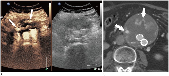
Abdominal aortic aneurysm (AAA) can be defined as an abnormal, progressive dilatation of the abdominal aorta, carrying a substantial risk for fatal aneurysmal rupture. Endovascular aneurysmal repair (EVAR) for AAA is a minimally invasive endovascular procedure that involves the placement of a bifurcated or tubular stent-graft over the AAA to exclude the aneurysm from arterial circulation. In contrast to open surgical repair, EVAR only requires a stab incision, shorter procedure time, and early recovery. Although EVAR seems to be an attractive solution with many advantages for AAA repair, there are detailed requirements and many important aspects should be understood before the procedure. In this comprehensive review, fundamental information regarding AAA and EVAR is presented.

Keywords: Abdominal aortic aneurysm; Endovascular aneurysmal repair; Stent-graft.


Conflict of interest statement

The authors have no potential conflicts of interest to disclose.

Figures

Fig. 1
Fig. 1. Aortic angle.
Angle between flow axes which are defined as central lumen lines of suprarenal and infrarenal aorta, forms aortic angle.
Fig. 2
Fig. 2. Shape of aneurysmal neck.
Aneurysmal neck can be straight (A), tapered (B), or reverse tapered (C). Tapered or reversed tapered neck is defined when there is difference more than 3 mm in diameter of proximal and distal aortic necks.
Fig. 3
Fig. 3. Various designs of stent-grafts.
A–C. Stent-grafts with suprarenal fixator. Fixating barbs are present on bare metallic stent portion around top of stent-graft. D. Stent-graft without fixating barbs as active sealing system. E. Stent with infrarenal fixator. Fixating barbs are located on graft fabric. A, B, E. Stent-grafts with exoskeleton design and metallic stents are mounted outside graft fabric. C, D. Stent-grafts with endoskeleton design and stents are mounted inside graft fabric.
Fig. 4
Fig. 4. Ultrasound-guided common femoral artery access.
Arterial wall puncture should be done as close to 12 o'clock direction of anterior wall (arrow) as possible to reduce access site complication. Note echogenic needle tip (arrowhead) within arterial lumen.
Fig. 5
Fig. 5. Lumbar artery embolization prior to EVAR to prevent type II endoleak.
A. Aortogram shows enlarged lumbar artery more than 2 mm in diameter (arrows). B. Radiograph obtained after EVAR. Embolization coils are placed in right lumbar artery (arrowheads). EVAR = endovascular aneurysmal repair
Fig. 6
Fig. 6. Aortogram should be obtained to localize orifices of bilateral renal arteries, prior to stent-graft deployment.
Changing C-arm angle to gather proximal markers onto virtual line (dashed line) as close as possible is helpful in assessing exact level of bilateral renal artery orifices (arrowheads).
Fig. 7
Fig. 7. Gate cannulation with snare technique.
A. Guidewire from ipsilateral access site is introduced into gate for contralateral limb (arrows), then is located around snare which introduced through contralateral vascular access site. B. After snaring, guidewire is externalized through contralateral vascular access site, then additional catheter is engaged along externalized guidewire.
Fig. 8
Fig. 8. External iliac artery rupture during EVAR.
A. Angiogram shows external iliac artery rupture during guidewire insertion and subsequent extravasation of contrast agent. B. Aortogram obtained after EVAR shows exclusion of external iliac artery rupture with limb extension to right external iliac artery.
Fig. 9
Fig. 9. External iliac artery dissection after EVAR.
A. Angiogram shows thin arterial dissection along left external iliac artery (arrows). B. Bare metal stent is additionally placed over external iliac artery dissection and then, intravascular flow is restored.
Fig. 10
Fig. 10. Right renal infarction after EVAR.
Contrast-enhanced CT acquired 4 days after EVAR shows right renal infarction with decreased renal perfusion.
Fig. 11
Fig. 11. Delayed stent-graft migration after successful EVAR.
A. Aortogram after EVAR shows exclusion of aneurysmal sac. Ends of fixating barbs are placed at proximal portion to both renal artery orifices (arrows). B. Aortogram obtained 4 years after EVAR demonstrates caudal migration of stent-graft. Note massive type Ia endoleak around proximal attachment site (arrowheads).
Fig. 12
Fig. 12. Diagram showing five different types of endoleaks.
Fig. 13
Fig. 13. Type Ib endoleak after EVAR.
A, B. Arteriograms acquired immediately after EVAR show massive type Ib endoleak from right side (arrows). C. After coil embolization of right internal iliac artery (arrows) and limb extension (arrowheads), type Ib endoleak disappeared.
Fig. 14
Fig. 14. Type II endoleak from lumbar artery after EVAR.
A. Arteriogram shows type II endoleak from internal iliac artery-lumbar artery collaterals (arrows). B. Follow-up non-contrast CT after embolization with glue, shows glue cast in endoleak nidus within aneurysmal sac and feeding lumbar artery (arrowheads).
Fig. 15
Fig. 15. Type IIIa endoleak due to modular disconnection.
A. Radiogram obtained few months after EVAR, shows modular disconnection between main body stent-graft and iliac limb extension stent-graft (arrows). Also note graft kinking at contralateral extension limb (arrowheads). B. Aortogram shows massive type IIIa endoleak (arrows) from modular disconnection site.
Fig. 16
Fig. 16. Delayed AAA rupture after type IIIa endoleak.
A. Non-contrast CT shows hyper-attenuating hematoma within aneurysmal sac and peri-aortic retroperitoneal space (arrows), suggesting delayed AAA rupture after EVAR. B. CT scan obtained at caudal level to A shows modular separation (arrowheads). AAA = abdominal aortic aneurysm
Fig. 17
Fig. 17. Limb occlusion after stent-graft kinking.
A. Aortogram shows absence of blood flow through right side limb at right iliac artery. Stent-graft kinking is also noted (arrow). B. Follow-up angiogram after stenting shows revascularization through right side limb stent-graft as well as improvement of stent-graft kinking (arrowheads).
Fig. 18
Fig. 18. Stent-graft infection.
A. Contrast-enhanced CT scan shows peri-graft fluid collection with enhancing wall (arrows). B. Photogram obtained after percutaneous aspiration shows pus-natured fluid.
Fig. 19
Fig. 19. Aorto-enteric fistula causing massive hematochezia in 45-year-old woman.
Contrast-enhanced CT scan shows stent-graft exposure (arrows) and extravasation of contrast agent into adjacent small bowel lumens (arrowheads).
Fig. 20
Fig. 20. EVAR under three-dimensional overlaid image.
After using simple software post-processing, pre-procedural CT data can be overlaid onto live intra-procedural fluoroscopic image, like roadmap image-guide.
Fig. 21
Fig. 21. CEUS for imaging surveillance after EVAR.
A. CEUS image shows contrast leakage within aneurysmal sac suggesting endoleak (arrows). B. Contrast-enhanced CT scan also demonstrates endoleak (arrows) in same patient. CEUS finding is well correlated with endoleak detected on contrast-enhanced CT. CEUS = contrast-enhanced ultrasound

Similar articles

Cited by

References

    1. Kuivaniemi H, Elmore JR. Opportunities in abdominal aortic aneurysm research: epidemiology, genetics, and pathophysiology. Ann Vasc Surg. 2012;26:862–870. - PubMed
    1. Wang LJ, Prabhakar AM, Kwolek CJ. Current status of the treatment of infrarenal abdominal aortic aneurysms. Cardiovasc Diagn Ther. 2018;8(Suppl 1):S191–S199. - PMC - PubMed
    1. Lederle FA, Johnson GR, Wilson SE, Ballard DJ, Jordan WD, Jr, Blebea J, et al. Veterans Affairs Cooperative Study #417 Investigators. Rupture rate of large abdominal aortic aneurysms in patients refusing or unfit for elective repair. JAMA. 2002;287:2968–2972. - PubMed
    1. Parodi JC, Palmaz JC, Barone HD. Transfemoral intraluminal graft implantation for abdominal aortic aneurysms. Ann Vasc Surg. 1991;5:491–499. - PubMed
    1. Schermerhorn ML, Bensley RP, Giles KA, Hurks R, O'malley AJ, Cotterill P, et al. Changes in abdominal aortic aneurysm rupture and short-term mortality, 1995-2008: a retrospective observational study. Ann Surg. 2012;256:651–658. - PMC - PubMed