

SQSTM1/p62 promotes mitochondrial ubiquitination independently of PINK1 and PRKN/parkin in mitophagy
- PMID: 31339428
- PMCID: PMC6844492
- DOI: 10.1080/15548627.2019.1643185
SQSTM1/p62 promotes mitochondrial ubiquitination independently of PINK1 and PRKN/parkin in mitophagy
Abstract
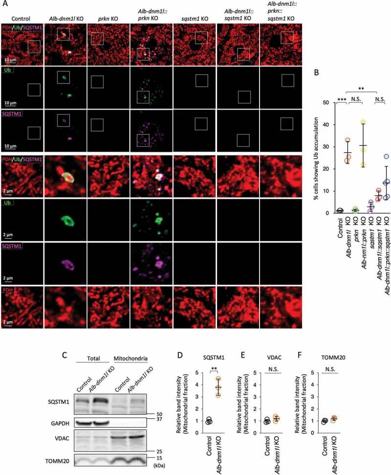
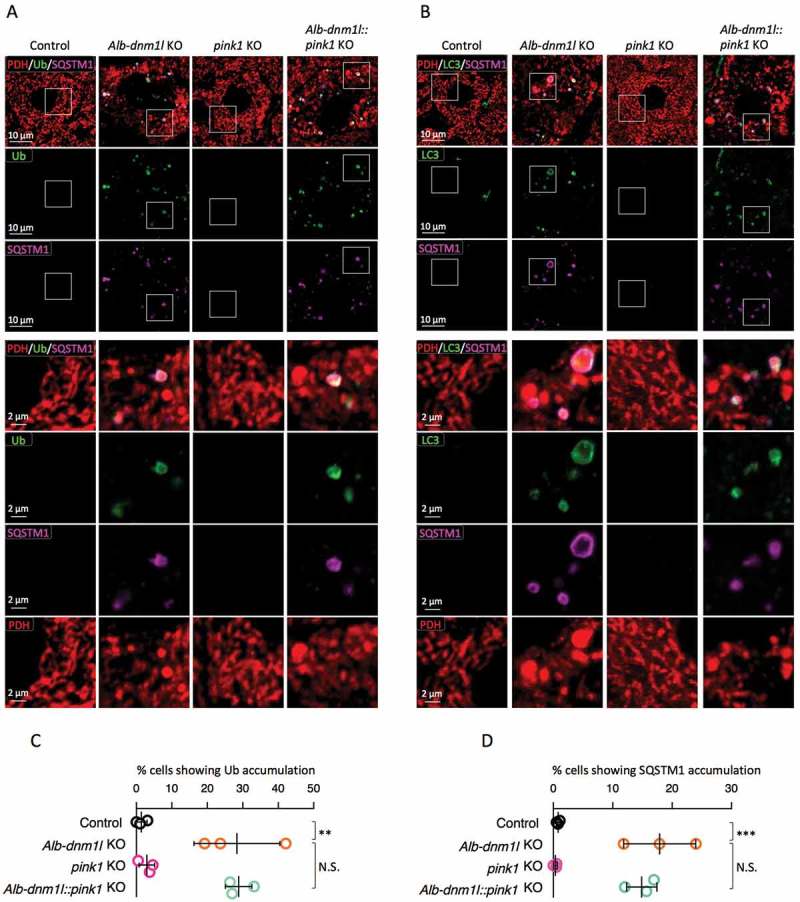
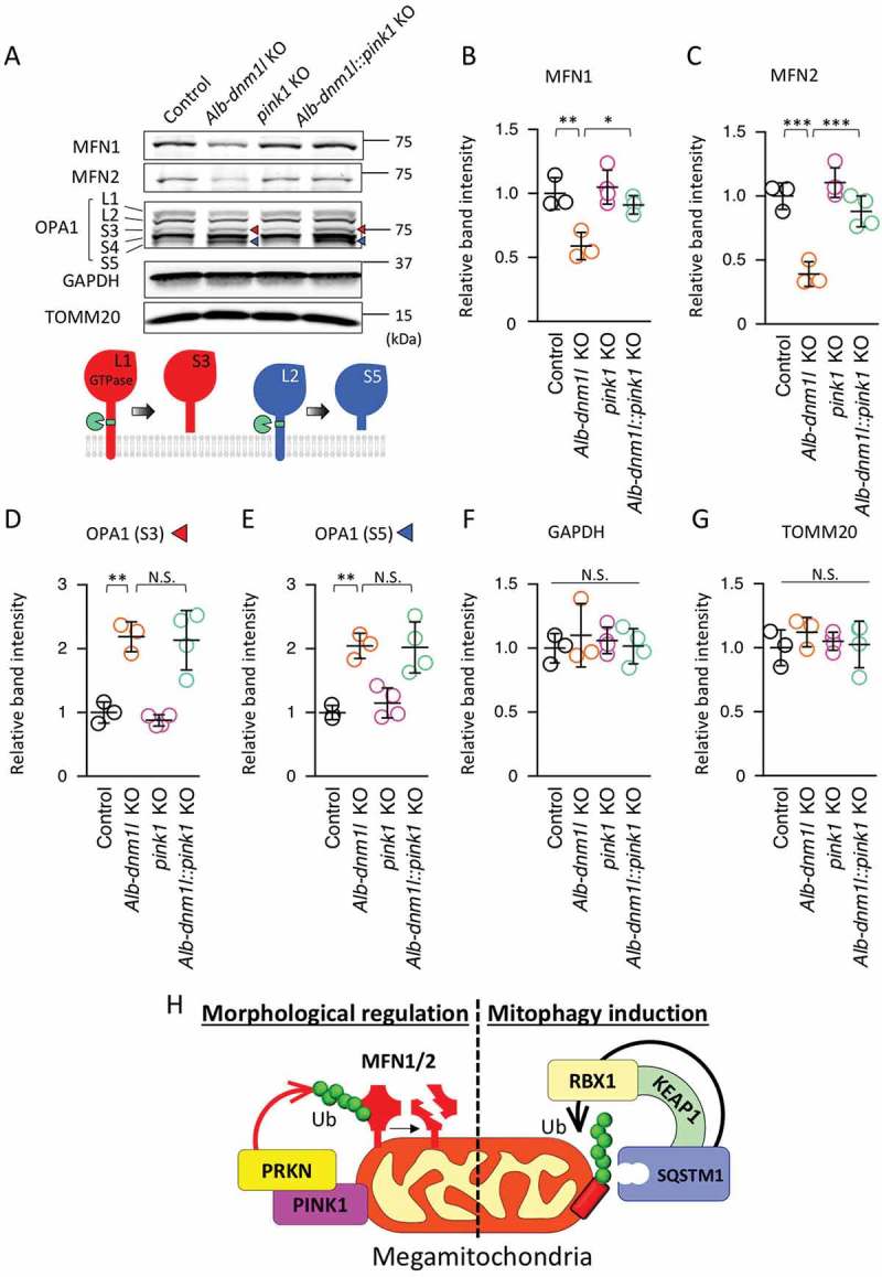
The ubiquitination of mitochondrial proteins labels damaged mitochondria for degradation through mitophagy. We recently developed an in vivo system in which mitophagy is slowed by inhibiting mitochondrial division through knockout of Dnm1l/Drp1, a dynamin related GTPase that mediates mitochondrial division. Using this system, we revealed that the ubiquitination of mitochondrial proteins required SQSTM1/p62, but not the ubiquitin E3 ligase PRKN/parkin, during mitophagy. Here, we tested the role of PINK1, a mitochondrial protein kinase that activates mitophagy by phosphorylating ubiquitin, in mitochondrial ubiquitination by knocking out Pink1 in dnm1l-knockout liver. We found mitochondrial ubiquitination did not decrease in the absence of PINK1; instead, PINK1 was required for the degradation of MFN1 (mitofusin 1) and MFN2, two homologous outer membrane proteins that mediate mitochondrial fusion in dnm1l-knockout hepatocytes. These data suggest that mitochondrial ubiquitination is promoted by SQSTM1 independently of PINK1 and PRKN during mitophagy. PINK1 and PRKN appear to control the balance between mitochondrial division and fusion in vivo. Abbreviations: DNM1L/DRP1: dynamin 1-like; KEAP1: kelch-like ECH-associated protein 1; KO: knockout; MAP1LC3/LC3: microtubule-associated protein 1 light chain 3; MFN1/2: mitofusin 1/2; OPA1: OPA1, mitochondrial dynamin like GTPase; PDH: pyruvate dehydrogenase E1; PINK1: PTEN induced putative kinase 1; PRKN/parkin: parkin RBR E3 ubiquitin protein ligase.
Keywords: Dnm1l/Drp1; PINK1; PRKN/parkin; mitochondria; mitochondrial division; mitophagy.
Figures
References
-
- Quiros PM, Langer T, Lopez-Otin C.. New roles for mitochondrial proteases in health, ageing and disease. Nat Rev Mol Cell Biol. 2015. June;16(6):345–359. .PubMed PMID: 25970558 - PubMed
-
- Goard CA, Schimmer AD. Mitochondrial matrix proteases as novel therapeutic targets in malignancy. Oncogene. 2014. May 22;33(21):2690–2699. . PubMed PMID: 23770858. - PubMed
-
- Shpilka T, Haynes CM. The mitochondrial UPR: mechanisms, physiological functions and implications in ageing. Nat Rev Mol Cell Biol. 2018. February;19(2):109–120. .PubMed PMID: 29165426 - PubMed
-
- Quiros PM, Mottis A, Auwerx J. Mitonuclear communication in homeostasis and stress. Nat Rev Mol Cell Biol. 2016. April;17(4):213–226. .PubMed PMID: 26956194 - PubMed
Publication types
MeSH terms
Substances
Grants and funding
LinkOut - more resources
Full Text Sources
Molecular Biology Databases
Research Materials
Miscellaneous