. 2019 Sep 26;47(17):9160-9179.
doi: 10.1093/nar/gkz639.

CTCF cooperates with CtIP to drive homologous recombination repair of double-strand breaks

Affiliations

CTCF cooperates with CtIP to drive homologous recombination repair of double-strand breaks

Soon Young Hwang et al. Nucleic Acids Res. .

Abstract

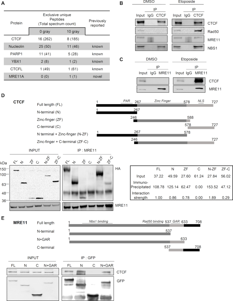
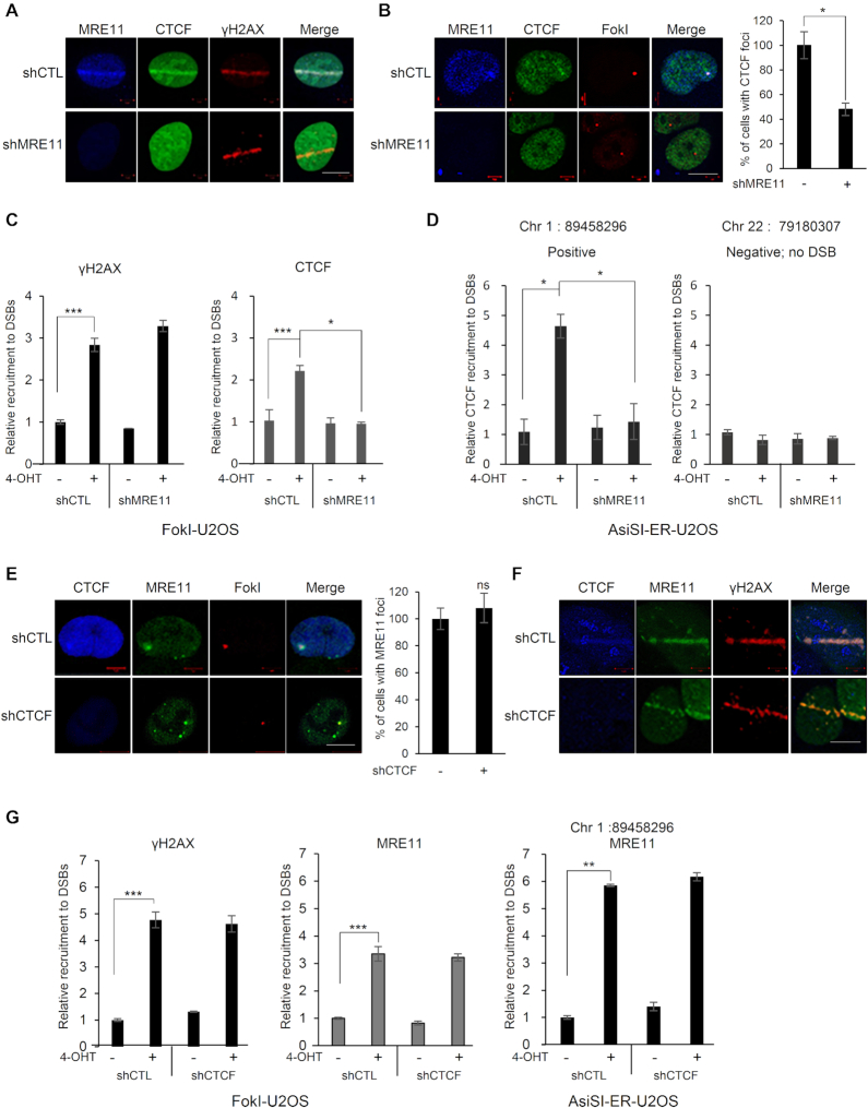
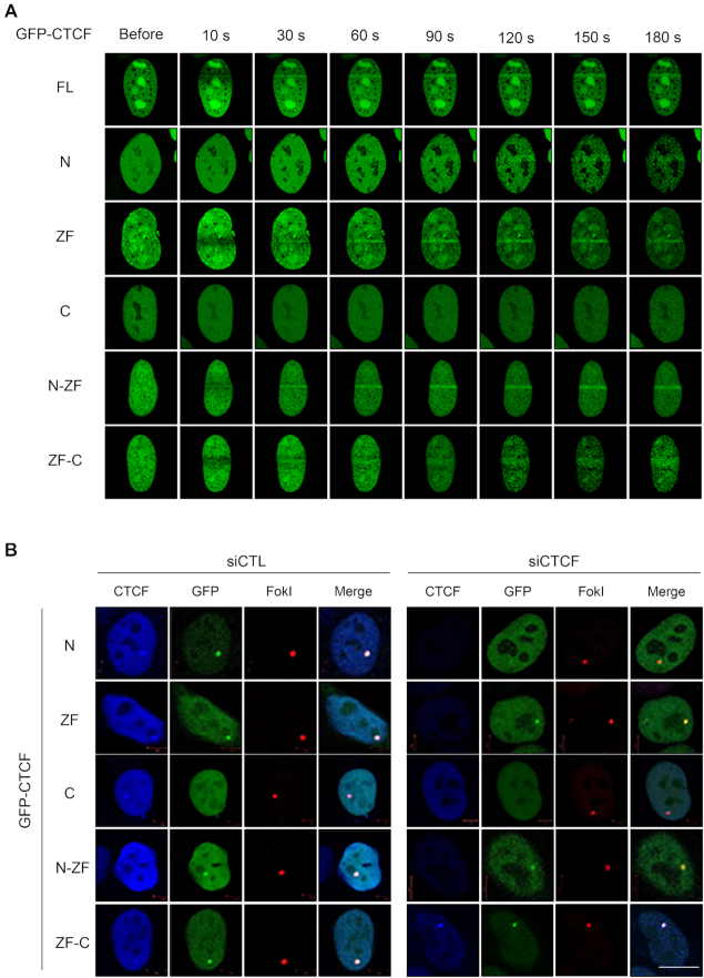
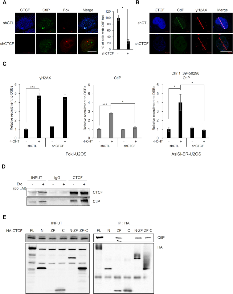
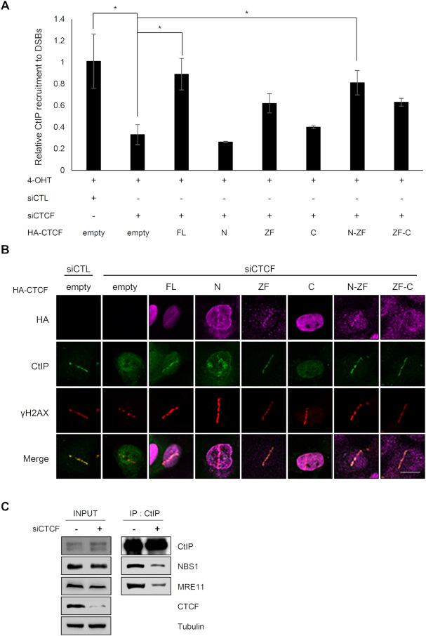
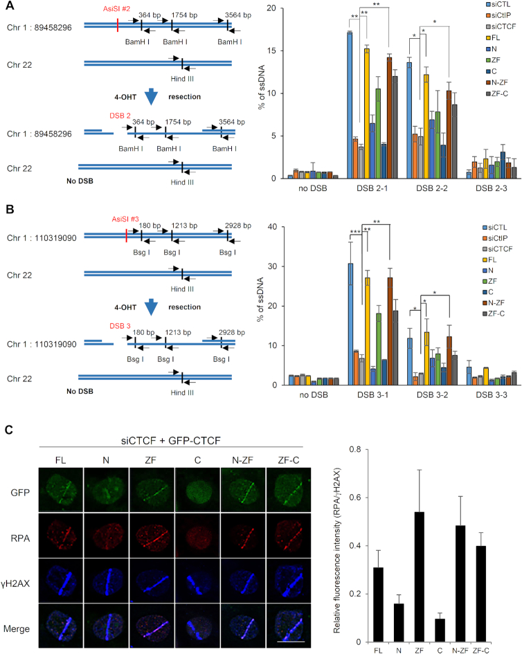
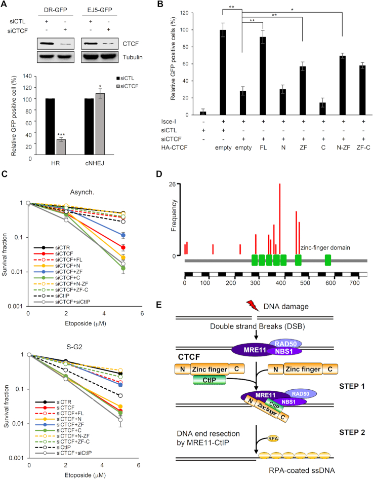
The pleiotropic CCCTC-binding factor (CTCF) plays a role in homologous recombination (HR) repair of DNA double-strand breaks (DSBs). However, the precise mechanistic role of CTCF in HR remains largely unclear. Here, we show that CTCF engages in DNA end resection, which is the initial, crucial step in HR, through its interactions with MRE11 and CtIP. Depletion of CTCF profoundly impairs HR and attenuates CtIP recruitment at DSBs. CTCF physically interacts with MRE11 and CtIP and promotes CtIP recruitment to sites of DNA damage. Subsequently, CTCF facilitates DNA end resection to allow HR, in conjunction with MRE11-CtIP. Notably, the zinc finger domain of CTCF binds to both MRE11 and CtIP and enables proficient CtIP recruitment, DNA end resection and HR. The N-terminus of CTCF is able to bind to only MRE11 and its C-terminus is incapable of binding to MRE11 and CtIP, thereby resulting in compromised CtIP recruitment, DSB resection and HR. Overall, this suggests an important function of CTCF in DNA end resection through the recruitment of CtIP at DSBs. Collectively, our findings identify a critical role of CTCF at the first control point in selecting the HR repair pathway.


Figures

Figure 1.
Figure 1.
CTCF interacts with MRE11 in response to DNA damage. (A) Selected protein list obtained from LC-MS/MS analysis after interactome tandem affinity purification of FLAG-SFB-tagged CTCF with or without γ-irradiation. The previously known and novel hits from MS results are shown. (B and C) Forward (B) and reciprocal (C) co-immunoprecipitation (co-IP) between endogenous CTCF and MRE11 in 293T cells, after the addition of DNase Benzonase, was performed with anti-CTCF (B) or anti-MRE11 (C) antibody, without (left, DMSO) and with (right, Etoposide) etoposide treatment. Immunoblot (IB) analysis was performed with the indicated antibodies. IgG immunoprecipitation (IP) was used as a negative control. See also Supplementary Figure S1. (D and E) Schematic representations of CTCF (D) and MRE11 (E) constructs used in this study (Top). The 293T cells were transfected with the indicated HA-tagged CTCF truncation constructs (D) or GFP-tagged MRE11 truncation constructs (E). Cell lysates were immunoprecipitated with anti-MRE11 (D) or anti-GFP (E) antibody, and immunoblot (IB) analyses were performed with the indicated antibodies. (D) Positions of molecular weight makers are denoted on the left of the INPUT SDS-PAGE gel. The interaction strengths between MRE11-HA and fusion CTCF proteins were normalized to the expression levels of HA fusion CTCF proteins, i.e. input. The interaction strength between MRE11-HA full-length CTCF was set as one, and the interaction strength of each HA fusion CTCF protein was calculated (bottom right panel); PAR, poly ADP-ribosylation; NLS, nuclear localization signal; N+GAR, N-terminus and glycine arginine rich motif.
Figure 2.
Figure 2.
MRE11-dependent recruitment of CTCF at sites of DNA damage. (A) MRE11-depleted (shMRE11) or control (shCTL) U2OS cells were presensitized with BrdU and subjected to laser micro-irradiation. Cells were fixed and stained with the indicated antibodies; scale bar: 10 μm. (B) Immunofluorescence was performed 4 h after induction of double-strand breaks by ER-mCherry-lacR-FokI-DD in the FokI-U2OS reporter cells transfected with shRNA targeting MRE11 (shMRE11) or control shRNA (shCTL); scale bar: 10 μm. The plot represents the percentage of cells positive for CTCF co-localized at mCherry-FokI foci. Data are the means ± SD of three independent experiments. More than 100 cells were counted in each experiment; *P≤ 0.05. (C) ChIP-qPCR was performed with an antibody to γ-H2AX or CTCF in the FokI-U2OS DSB reporter cells transfected with the indicated shRNA (shCTL as a control or shMRE11 targeting MRE11), with (+) or without (−) induction of DSBs by mCherry-LacI-FokI. The values of recruitment to DSBs were relative to those of cells without the induction of DSBs. All qPCR reactions were performed in triplicate, with the SEM values calculated from at least three independent experiments; *P≤ 0.05; ***P≤ 0.001. (D) AsiSI-ER-U2OS cells were transfected with the indicated shRNA, with (+) or without (−) induction of DSBs by AsiSI. CTCF and chromatin were immunoprecipitated with anti-CTCF antibody. The fold enrichment values were relative to those of cells without induction of DSBs. Primers on chromosome 22 (no DSB) were used as negative controls. Data are the means ± SD of at least three independent experiments, and all qPCR reactions were performed in triplicate. (E) Immunofluorescence was performed 4 h after induction of double-strand breaks (DSBs) by mCherry-LacI-FokI in the CTCF-depleted (shCTCF) or control (shCTL) FokI-U2OS cells; scale bar: 10 μm. Bar graph represents the percentage of cells positive for MRE11 co-localized at mCherry-LacI-FokI foci. Data are the means ± SD of three independent experiments. More than 100 cells were counted in each experiment. ns, not significant. (F) Recruitment of MRE11 (green) to DSBs induced by laser micro-irradiation in the CTCF-depleted (shCTCF) and control (shCTL) U2OS cells; scale bar: 10 μm. (G) ChIP-qPCR was performed with an antibody to γ-H2AX or MRE11 in FokI-U2OS cells (left and center), and AsiSI-ER-U2OS cells (right) transfected with the control (shCTL) or CTCF shRNA (shCTCF), with (+) or without (−) induction of DSBs by FokI (FokI-U2OS) or AsiSI (AsiSI-ER-U2OS). The fold enrichment values were relative to those of cells without induction of DSBs. Data are presented as means ± SD of three independent experiments, and all qPCR reactions were performed in triplicate; **P≤ 0.01; ***P≤ 0.001.
Figure 3.
Figure 3.
CTCF recruitment to DNA lesions requires its N-terminal or zinc-finger domain. (A) U2OS cells were transfected with GFP-tagged full-length CTCF and its truncation constructs (see Figure 1D, top), and were subjected to laser micro-irradiation for up to 180 s. (B) Recruitment of the indicated GFP-tagged CTCF proteins (green; see Figure 1D top) to double-strand breaks induced by mCherry-LacI-FokI (red) in the endogenous CTCF-depleted (siCTCF) or control (siCTL) FokI-U2OS cells. The ectopically complemented C-terminal domain of CTCF in CTCF knockdown cells (GFP-tagged C and ZF-C in siCTCF cells, right) is also recognized with an antibody to endogenous CTCF (blue); scale bar: 10 μm.
Figure 4.
Figure 4.
CTCF is required for CtIP recruitment at DNA lesions. (A) Immunofluorescence was performed 4 h after induction of double-strand breaks (DSBs) by mCherry-LacI-FokI in CTCF-depleted (shCTCF) or control (shCTL) FokI-U2OS cells; scale bar: 10 μm. Bar graph represents the percentage of cells with CtIP (green) that co-localized at mCherry-FokI (red) foci. Data are the means ± SD of at least three independent experiments. More than 100 cells were counted in each experiment; *P≤ 0.05. (B) CTCF-depleted (shCTCF) or control (shCTL) U2OS cells were subjected to laser micro-irradiation. Cells were fixed and stained with the indicated antibodies. The scale bar represents 10 μm. (C) ChIP-qPCR was performed with an antibody to CtIP and γ-H2AX in the CTCF-depleted (shCTCF) or control (shCTL) FokI-U2OS cells (left) and AsiSI-ER-U2OS cells (right), with (+) or without (−) induction of DSBs. The fold enrichment values were relative to those of cells without induction of DSBs. Data are means ± SD of at least three independent experiments, and all qPCR reactions were performed in triplicate; *P≤ 0.05; ***P≤ 0.001. (D) CTCF interacts with CtIP. Co-immunoprecipitation assays were performed using control IgG and anti-CTCF with (+) or without (−) etoposide (Eto) treatment, and the immunoprecipitates were probed for the indicated proteins by western blotting (IB). (E) CTCF interacts with CtIP via its zinc finger domain. HA-tagged full-length CTCF and its truncated fragments (see Figure 1D, top) were expressed in 293T cells. HA-immunoprecipitates were resolved by SDS-PAGE, and blots were probed for HA (bottom) and CtIP (top).
Figure 5.
Figure 5.
CtIP recruitment to DNA damage requires interactions with the zinc finger domain of CTCF. (A) Recruitment of CtIP to mCherry-LacI-FokI-induced DSBs was evaluated by ChIP–qPCR in the CTCF-depleted FokI–U2OS cells complemented by the indicated HA fusion CTCF proteins (see Figure 1D, top). CtIP and chromatin were immunoprecipitated with an anti-CtIP antibody. qPCR was performed for the quantitative analysis of ChIP samples. Fold recruitment values were relative to those of cells without induction of DSBs. Data are means ± SD of at least three independent experiments, and all qPCR reactions were performed in triplicate; *P≤ 0.05. (B) GFP–CtIP co-localization (green) at γH2AX foci (red) in CTCF-depleted U2OS cells complemented by the indicated HA fusion CTCF proteins (see Figure 1D, top). CTCF-depleted (siCTCF) and control (siCTL) U2OS cells were subjected to laser micro-irradiation. Cells were fixed and stained with the antibodies indicated on the left. The scale bar represents 10 μm. (C) Co-immunoprecipitation of endogenous CtIP and MRE11 in CTCF-depleted (+) or control (−) 293T cells was performed with anti-CtIP antibody. Immunoblot (IB) analysis was performed with the antibodies indicated on the right.
Figure 6.
Figure 6.
CTCF depletion impairs DNA end resection. (A) Quantification of ssDNA generated by 5′-end resection at three sites (left, 2-1, 2-2, and 2–3 are 364, 1754 and 3564 bp from DSB, respectively and the paired primers across BamH 1 restriction sites are indicated as black arrow pairs) around the AsiSI-induced DSB (red) on chromosome 1:89,458,296 position in AsiSI–ER–U2OS cells transfected with the indicated shRNAs and HA-tagged CTCF constructs (right plot). The primers (black arrow pair) on chromosome 22 (no DSB) across a Hind III restriction site were used for negative control (left). Data are means ± SD of at least three independent experiments, and all qPCR reactions were performed in triplicate; *P≤ 0.05, **P≤ 0.01 (see Supplementary Figure S13A indicating levels of depleted CTCF or CtIP and complemented HA-tagged CTCF proteins.). (B) Design of qPCR primers and probes for measurement of resection at sites, 3-1 (180 bp), 3-2 (1213 bp) and 3-3 (2928 bp) near an AsiSI-induced DSB (red) on chromosome 1:110,319,090 position (left). Quantification of ssDNA generated from resection at three sites (left, 3-1, 3-2 and 3-3 located at Bsg I restriction sites) as in part (A) (right plot). Data represent means ± SEM of three independent experiments (see Supplementary Figure S13B indicating levels of silenced and complemented proteins.). (C) Accumulation of RPA onto the laser strips in CTCF-depleted U2OS cells complemented with the indicated GFP-tagged full-length or truncated CTCF proteins (left, see Figure 1A, above). Relative fluorescent intensities of RPA to those of γ-H2AX are presented in the right plot as the means ± SD of at least three independent experiments. More than 30 cells were counted in each experiment.
Figure 7.
Figure 7.
CTCF–CtIP interaction contributes to CTCF-mediated homologous recombination (HR) and survival upon exposure to DNA damage. (A) U2OS-based homologous recombination- (DR-GFP) or canonical non-homologous end joining- (EJ5-GFP) reporter cell lines were co-transfected with siRNA (control, siCTL, or endogenous CTCF-specific, siCTCF) and I-SceI as indicated. Homologous recombination (HR) and canonical non-homologous end joining (cNHEJ) efficiencies were analyzed as in part (A). The plotted values are means ± SEM from at least three independent experiments; *P≤ 0.05; ***P≤ 0.001. (B) HR reporter cells (DR-GFP) were transfected with the indicated siRNAs and HA-tagged CTCF constructs together with I-SceI and subjected to an HR assay as in part (A) (see Supplementary Figure S13C indicating knockdown of endogenous CTCF and supplementation with the full-length or truncated constructs of CTCF.) The plotted values are means ± SEM from at least three independent experiments; *P≤ 0.05; **P≤ 0.01. (C) Effect of ectopic expression of the indicated HA-tagged CTCF proteins on survival of CTCF-depleted HeLa cells after treatment with etoposide. HeLa cells were transfected with the indicated siRNAs and HA-tagged CTCF constructs. The cells in asynchronous or S/G2 phase were treated with etoposide, and survival fractions were confirmed by a clonogenic survival assay. The intensities and areas of the colonies were measured using ImageJ software (see Supplementary Figure S13D showing depletion of CTCF or CtIP and supplemented CTCF protein levels.). (D) Somatic point mutations (nonsense and missense) of CTCF from Catalogue of Somatic Mutations in Cancer (COSMIC) as of 13 October, 2018. Among them, 14 substitution mutations were identified in ≥4 samples across tumors in the COSMIC database. Eight out of the 14 mutations were located on the zinc finger (ZF) domain, which was defined as Pfam (ID P49711). As the frequency of mutations in the ZF domain of CTCF was higher than that in its N-terminal domain, the somatic mutations were significantly enriched within the ZF domain in cancer. (E) Model for the role of CTCF in DNA end resection of HR-mediated repair via CtIP recruitment. The MRN complex recognizes and binds to double-strand breaks (DSBs) and recruits CTCF to sites of DNA damage. In turn, CTCF promotes the recruitment of CtIP to DSBs and enhances DNA end resection.

References

    1. Jimeno S., Femandez-Avila M.J., Cruz-Garcia A., Cepeda-Garcia C., Gomez-Cabello D., Huertas P.. Neddylation inhibits CtIP-mediated resection and regulates DNA double strand break repair pathway choice. Nucleic Acids Res. 2015; 43:987–999. - PMC - PubMed
    1. Yuan J., Chen J.. N terminus of CtIP is critical for homologous recombination-mediated double-strand break repair. J. Biol. Chem. 2009; 284:31746–31752. - PMC - PubMed
    1. Huertas P. DNA resection in eukaryotes: deciding how to fix the break. Nat. Struct. Mol. Biol. 2010; 17:11–16. - PMC - PubMed
    1. Forget A.L., Kowalczykowski S.C.. Single-molecule imaging brings Rad51 nucleoprotein filaments into focus. Trends Cell Biol. 2010; 20:269–276. - PMC - PubMed
    1. San Filippo J., Sung P., Klein H.. Mechanism of eukaryotic homologous recombination. Annu. Rev. Biochem. 2008; 77:229–257. - PubMed

Publication types