Review
. 2019 Jul 23;20(14):3590.
doi: 10.3390/ijms20143590.

Molecular Mechanisms of p63-Mediated Squamous Cancer Pathogenesis

Affiliations
Review

Molecular Mechanisms of p63-Mediated Squamous Cancer Pathogenesis

Michael A Moses et al. Int J Mol Sci. .

Abstract

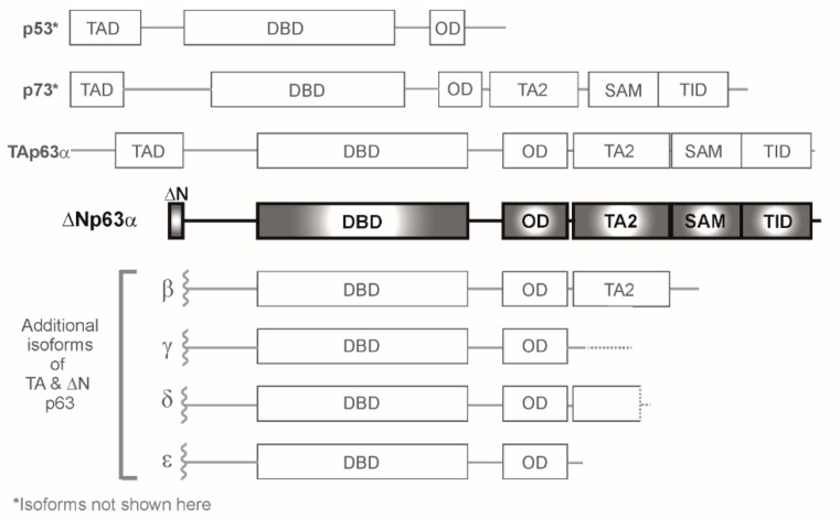
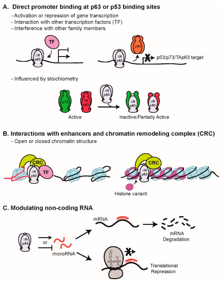
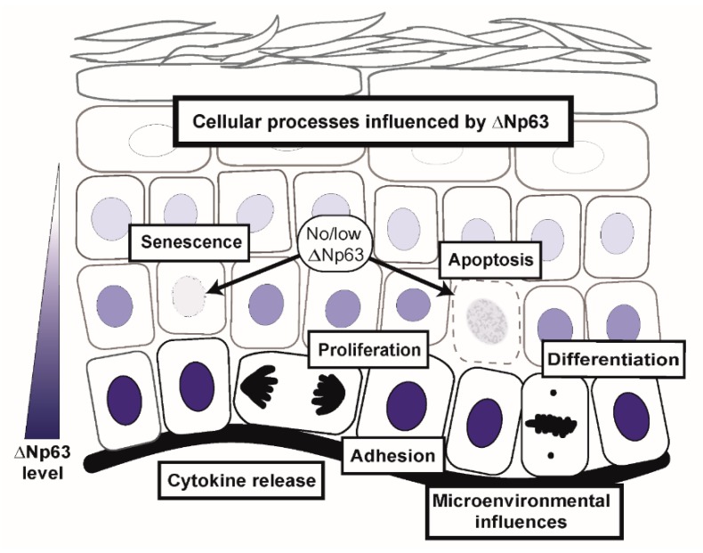
The p63 gene is a member of the p53/p63/p73 family of transcription factors and plays a critical role in development and homeostasis of squamous epithelium. p63 is transcribed as multiple isoforms; ΔNp63α, the predominant p63 isoform in stratified squamous epithelium, is localized to the basal cells and is overexpressed in squamous cell cancers of multiple organ sites, including skin, head and neck, and lung. Further, p63 is considered a stem cell marker, and within the epidermis, ΔNp63α directs lineage commitment. ΔNp63α has been implicated in numerous processes of skin biology that impact normal epidermal homeostasis and can contribute to squamous cancer pathogenesis by supporting proliferation and survival with roles in blocking terminal differentiation, apoptosis, and senescence, and influencing adhesion and migration. ΔNp63α overexpression may also influence the tissue microenvironment through remodeling of the extracellular matrix and vasculature, as well as by enhancing cytokine and chemokine secretion to recruit pro-inflammatory infiltrate. This review focuses on the role of ΔNp63α in normal epidermal biology and how dysregulation can contribute to cutaneous squamous cancer development, drawing from knowledge also gained by squamous cancers from other organ sites that share p63 overexpression as a defining feature.

Keywords: epidermal homeostasis; epidermal morphogenesis; keratinocytes; p53 family; p63; squamous carcinogenesis.


Conflict of interest statement

The authors declare no conflicts of interest.

Figures

Figure 1
Figure 1
Schematic of p53/p63/p73 family members. Within the p63 homologues, alternative promoter usage yields TAp63 and ΔNp63 subclasses; within each subclass, alternative spicing yields α, β, γ, δ, and ε isoforms. This review focuses on ΔNp63α (highlighted), the predominant isoform in squamous epithelium. Domain abbreviations: TAD—Transactivation domain, ΔN—Delta N Domain, DBD—DNA binding domain, OD—Oligomerization domain, TA2—Transactivation domain 2, SAM—Sterile alpha motif, TID—Transactivation-inhibitory domain.
Figure 2
Figure 2
p63 mediates gene expression via multiple mechanisms. (A) Direct promoter binding: ΔNp63α activates or represses transcription by binding to promoters of its target genes and associating with other transcription factors to enhance or suppress transcriptional activity. The stoichiometric balance of the p63 and p73 isoforms can modulate the activity; these interactions can lead to different transcriptional programs and altered biological outcomes. (B) Interactions with enhancers and chromatin remodeling complex: At sites of high histone acetylation (e.g.; H3K27), p63 interaction with the chromatin remodeling complex (CRC) maintains an open chromatin landscape controlling access to epidermal enhancers and super enhancers to drive gene activation in conjunction with other transcription factors. In this way, p63 directs tissue-specific gene regulation guiding epidermal differentiation. (C) Activation or repression of non-coding RNAs: Long non-coding RNA and microRNA are targets of ΔNp63 regulation, thereby indirectly modulating gene expression. Examples of ΔNp63α activating or repressing transcription of miRNAs are shown.
Figure 3
Figure 3
ΔNp63α regulates cell intrinsic and extrinsic biological processes involved in normal epidermal morphogenesis and homeostasis. In cancer, amplification of p63 leads to the hijacking of these processes to support conversion to and progression of the malignant state.

References

    1. Yang A., Kaghad M., Wang Y., Gillett E., Fleming M.D., Dotsch V., Andrews N.C., Caput D., McKeon F. p63, a p53 homolog at 3q27-29, encodes multiple products with transactivating, death-inducing, and dominant-negative activities. Mol. Cell. 1998;2:305–316. doi: 10.1016/S1097-2765(00)80275-0. - DOI - PubMed
    1. Kaghad M., Bonnet H., Yang A., Creancier L., Biscan J.C., Valent A., Minty A., Chalon P., Lelias J.M., Dumont X., et al. Monoallelically expressed gene related to p53 at 1p36, a region frequently deleted in neuroblastoma and other human cancers. Cell. 1997;90:809–819. doi: 10.1016/S0092-8674(00)80540-1. - DOI - PubMed
    1. Dohn M., Zhang S., Chen X. p63alpha and DeltaNp63alpha can induce cell cycle arrest and apoptosis and differentially regulate p53 target genes. Oncogene. 2001;20:3193–3205. doi: 10.1038/sj.onc.1204427. - DOI - PubMed
    1. Ghioni P., Bolognese F., Duijf P.H., Van Bokhoven H., Mantovani R., Guerrini L. Complex transcriptional effects of p63 isoforms: identification of novel activation and repression domains. Mol. Cell. Biol. 2002;22:8659–8668. doi: 10.1128/MCB.22.24.8659-8668.2002. - DOI - PMC - PubMed
    1. Tanaka Y., Kameoka M., Itaya A., Ota K., Yoshihara K. Regulation of HSF1-responsive gene expression by N-terminal truncated form of p73alpha. Biochem. Biophys. Res. Commun. 2004;317:865–872. doi: 10.1016/j.bbrc.2004.03.124. - DOI - PubMed

MeSH terms