Review
. 2019 Jul 23;11(7):1036.
doi: 10.3390/cancers11071036.

Morphology and Molecular Features of Rare Colorectal Carcinoma Histotypes

Affiliations
Review

Morphology and Molecular Features of Rare Colorectal Carcinoma Histotypes

Andrea Remo et al. Cancers (Basel). .

Abstract

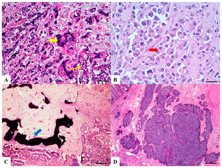
Several histopathological variants of colorectal carcinoma can be distinguished, some associated with specific molecular profiles. However, in routine practice, ninety/ninety-five percent of all large bowel tumors are diagnosed as conventional adenocarcinoma, even though they are a heterogeneous group including rare histotypes, which are often under-recognized. Indeed, colorectal cancer exhibits differences in incidence, location of tumor, pathogenesis, molecular pathways and outcome depending on histotype. The aim is therefore to review the morphological and molecular features of these rare variants of intestinal carcinomas which may hold the key to differences in prognosis and treatment.

Keywords: adenocarcinoma with osseous metaplasia; clear cell carcinoma; colorectal cancer histotypes; cribriform/comedo-type carcinoma; hepatoid carcinoma; lymphoepitelioma-like carcinoma; medullary carcinoma; micropapillary carcinoma; rhabdoid carcinoma; signet ring cell carcinoma.


Conflict of interest statement

The authors declare no conflict of interest.

Figures

Figure 1
Figure 1
Pie chart showing the frequency of colorectal carcinomas by histologic type.
Figure 2
Figure 2
Haematoxylin and Eosin stained sections of rare type colorectal carcinomas. (A) Serrated Adenocarcinoma: epithelial serrations or tufts (thick blue arrow), abundant eosinophilic or clear cytoplasm, vesicular basal nuclei with preserved polarity. Scale bar 200 micron. (B) Mucinous Carcinoma: presence of extracellular mucin (>50%) associated with ribbons or tubular structures of neoplastic epithelium. Scale bar 2 mm. (C) Signet Ring Carcinoma: more than 50% of signet cells with infiltrative growth pattern (thin red arrow) or floating in large pools of mucin (thick red arrow). Scale bar 200 micron. (D) Medullary carcinoma: neoplastic cells with syncytial appearance (thick yellow arrow) and eosinophilic cytoplasm associated with abundant peritumoral and intratumoral lymphocytes. Scale bar 100 micron.
Figure 3
Figure 3
Haematoxylin and Eosin stained sections of rare type colorectal carcinomas. (A) Lymphoepitelioma-like carcinoma: poorly differentiated cells (red arrow) arranged in solid nests, tubules and trabeculae with poorly demarcated, infiltrative margins; intratumoral lymphoid infiltrate is extremely abundant. Scale bar 200 micron. (B) Cribiform comedo-type carcinoma: cribriform gland (yellow arrow) with central necrosis comedo-like (yellow asterisk). Scale bar 400 micron. (C) Micropapillary Carcinoma: small, tight round to oval cohesive clusters of neoplastic cells (>5 cells) floating in clear spaces (double circle red-black), without endothelial lining and with no evidence of inflammatory cells. Scale bar 200 micron. (D) Low grade tubulo-glandular carcinoma: very well-differentiated invasive glands with uniform circular or tubular profiles (blue arrow) with bland cytologic atypia. Scale bar 400 micron.
Figure 4
Figure 4
Haematoxylin and Eosin stained sections of rare type colorectal carcinomas. (A) Villous carcinoma: invasive carcinoma with villous features consisting of usually intraglandular papillary projections (yellow arrow) associated with an expansile growth pattern, at the deep portions of the tumor. Scale bar 400 micron. (B) Squamous carcinoma: morphologically similar to other squamous cell carcinomas occurring in other organs with possible keratinization. Scale bar 200 micron. (C) Clear cell carcinoma: clear cell cytoplasm identified in polygonal cells with a central nucleus, columnar cells with an eccentric nucleus (red arrow) and/or round/oval cells with abundant cytoplasm and inconspicuous marginally located nucleus similar to lipocytes or lipoblasts. Scale bar 50 micron. (D) Hepatoid carcinoma: large polygonal-shaped cells, with granular eosinophilic cytoplasm, prominent nucleoli and trabecular and pseudo-acinar growth pattern similar to hepatocarcinoma. Scale bar 200 micron.
Figure 5
Figure 5
Haematoxylin and Eosin stained sections of rare type colorectal carcinomas. (A) Colorectal Choriocarcinoma: biphasic solid nests and trabeculae of mononucleated cells with clear cytoplasm (thin yellow arrow) and pleomorphic cells with abundant vacuolated or eosinophilic cytoplasm and single or multiple vescicular nuclei with conspicuous nucleoli (thick yellow arrow). Scale bar 200 micron. (B) Rhabdoid Colorectal Carcinoma: rhabdoid cells characterized by a large, eccentrically located nuclei, prominent nucleoli (red arrow) and abundant eosinophilic cytoplasm. Scale bar 100 micron. (C) Carcinoma with osseous metaplasia: osseous metaplasia (blue arrow) is recognized in conventional CRC as foci of bone formation in the stroma, with calcification, osteoid matrix, osteoclasts and osteoblasts. Scale bar 400 micron. (D) Undifferentiated carcinoma: sheets of undifferentiated cells showing a variable grade of pleomorphism with no gland formation, mucin production or other line of differentiation. Scale bar 400 micron.

References

    1. Remo A., Pancione M., Zanella C., Vendraminelli R. Molecular pathology of colorectal carcinoma. A systematic review centred on the new role of the pathologist. Pathologica. 2012;104:432–441. - PubMed
    1. Pancione M., Giordano G., Remo A., Febbraro A., Sabatino L., Manfrin E., Ceccarelli M., Colantuoni V. Immune escape mechanisms in colorectal cancer pathogenesis and liver metastasis. J. Immunol. Res. 2014;2014:686879. doi: 10.1155/2014/686879. - DOI - PMC - PubMed
    1. Pancione M., Remo A., Colantuoni V. Genetic and epigenetic events generate multiple pathways in colorectal cancer progression. Pathol. Res. Int. 2012;2012:509348. doi: 10.1155/2012/509348. - DOI - PMC - PubMed
    1. Shih I.M., Wang T.L., Traverso G., Romans K., Hamilton S.R., Ben-Sasson S., Kinzler K.W., Vogelstein B. Top-down morphogenesis of colorectal tumors. Proc. Natl. Acad. Sci. USA. 2001;98:2640–2645. doi: 10.1073/pnas.051629398. - DOI - PMC - PubMed
    1. Young J., Jenkins M., Parry S., Young B., Nancarrow D., English D., Giles G., Jass J. Serrated pathway colorectal cancer in the population: Genetic consideration. Gut. 2007;56:1453–1459. doi: 10.1136/gut.2007.126870. - DOI - PMC - PubMed

LinkOut - more resources