Review
. 2019 Jul 23;20(14):3598.
doi: 10.3390/ijms20143598.

Cell Death in the Kidney

Affiliations
Review

Cell Death in the Kidney

Giovanna Priante et al. Int J Mol Sci. .

Abstract

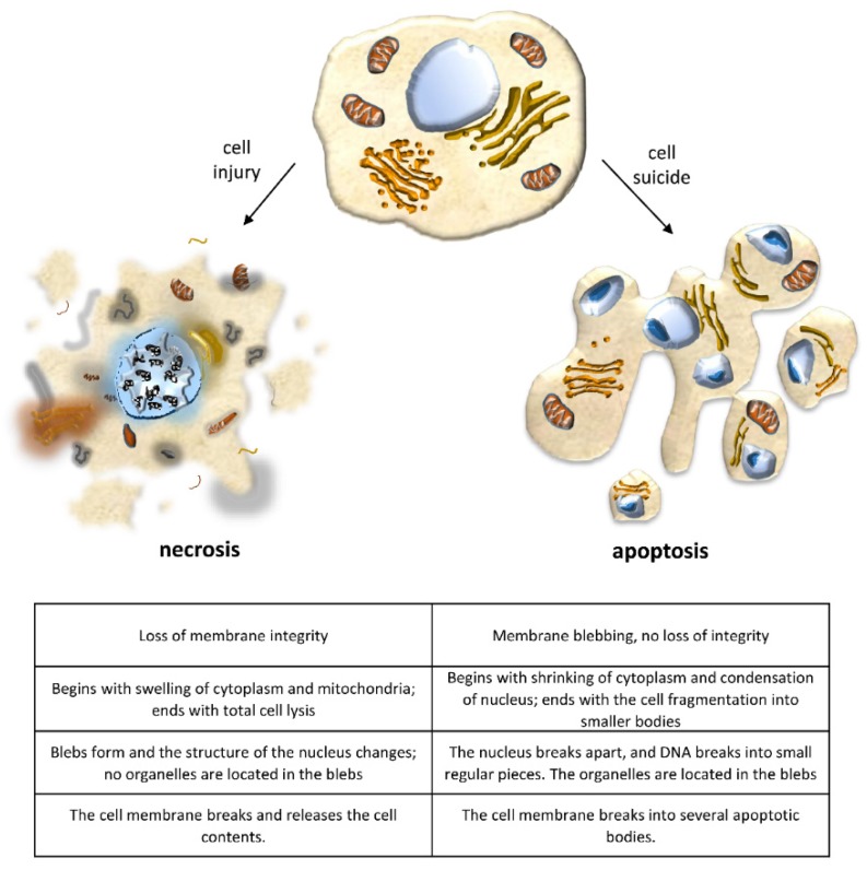
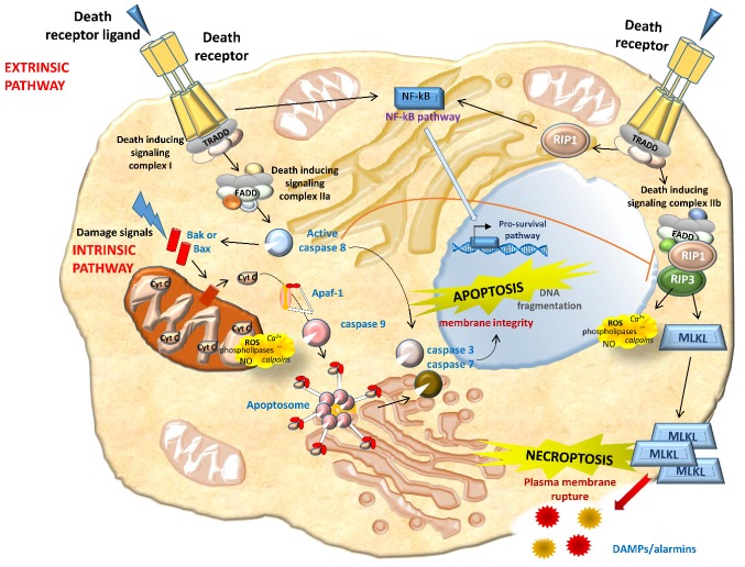
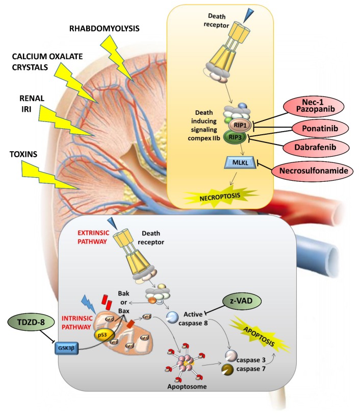
Apoptotic cell death is usually a response to the cell's microenvironment. In the kidney, apoptosis contributes to parenchymal cell loss in the course of acute and chronic renal injury, but does not trigger an inflammatory response. What distinguishes necrosis from apoptosis is the rupture of the plasma membrane, so necrotic cell death is accompanied by the release of unprocessed intracellular content, including cellular organelles, which are highly immunogenic proteins. The relative contribution of apoptosis and necrosis to injury varies, depending on the severity of the insult. Regulated cell death may result from immunologically silent apoptosis or from immunogenic necrosis. Recent advances have enhanced the most revolutionary concept of regulated necrosis. Several modalities of regulated necrosis have been described, such as necroptosis, ferroptosis, pyroptosis, and mitochondrial permeability transition-dependent regulated necrosis. We review the different modalities of apoptosis, necrosis, and regulated necrosis in kidney injury, focusing particularly on evidence implicating cell death in ectopic renal calcification. We also review the evidence for the role of cell death in kidney injury, which may pave the way for new therapeutic opportunities.

Keywords: apoptosis; glomerular injury; kidney injury; necrosis; regulated necrosis; tubular injury.


Conflict of interest statement

The authors have no conflicts of interest to disclose.

Figures

Figure 1
Figure 1
Changes in cell morphology that distinguish apoptosis from necrosis.
Figure 2
Figure 2
Overview of key molecular pathways of apoptosis and necroptosis. Apoptosis can start via intrinsic pathways (perturbation of intracellular homeostasis) or extrinsic pathways (death receptor binding). In the former case, cell stress leads directly (or via mediators, such as Bax and Bak) to mitochondrial outer membrane permeabilization, resulting in the release of apoptogenic factors, including Cytochrome c, which binds Apaf-1 to stimulate caspase-9 via apoptosomes. Bcl-2-related proteins induce apoptosis (e.g., Bax) or protect against it (e.g., Bcl-2). As for the extrinsic pathway, death receptor binding guides the recruitment of adapter proteins such as TRADD (TNFR-associated death domain), forming complex I. While complex I promotes cell survival via NF-кB activation, its transition to a secondary cytosolic complex, complex II, mediates cell death. Complex II is formed through the association of complex I with FADD (Fas-associated death domain). The formation of complex IIa promotes the activation of apoptosis in a caspase-8-dependent manner. Upon inhibition of caspase 8, complex IIb promotes necroptosis. Caspase-8 or caspase-9 activation subsequently triggers executioner caspases, such as caspase-3, -6, and -7. The cleavage of receptor-interacting protein kinase (RIPK) 1 and 3 by caspase-8 leads to apoptosis, whereas their phosphorylation triggers necroptosis in conditions of caspase-8 inhibition. RIPK1 and RIPK3 activation in turn causes the recruitment of the executioner mixed-lineage kinase domain-like protein (MLKL), which is phosphorylated by RIPK3 and initiates structural changes, leading to its insertion into the plasma membrane and channel formation. MLK channels increase Na+ influx, osmotic pressure, and membrane rupture, ending in cell death. Membrane rupture promotes the release of intracellular contents and endogenous damage-associated molecular patterns (DAMPs) and/or preformed proinflammatory molecules (alarmins). Through RIPK1 kinase activity, a wide range of necrotic mediators are activated in the execution phase of necrotic cell death, including reactive oxygen species (ROS), calcium (Ca2+), calpains, cathepsins, phospholipases, and ceramide.
Figure 3
Figure 3
Targeting apoptosis and necroptosis in kidney lesions. Different kidney lesions activate different cell death modalities: z-VAD (z-Val-Ala-Asp fluoromethyl ketone) has been found to protect renal function, and the effect of the pan-caspase inhibitor z-VAD on experimental renal ischemia-reperfusion (I/R) injury was to reduce serum urea levels, thereby preventing inflammation. TDZD-8 (4-benzyl-2-methyl-1,2,4-thiadiazolidine-3,5-dione) has been found to inhibit ischemia-induced activation of GSK3β (glycogen synthase kinase 3 beta), Bax, and caspase 3, thus ameliorating tubular and epithelial cell damage and significantly protecting renal function.

References

    1. Bessis M. Studies on cell agony and death: An attempt at classification. In: de Reuck A.V.S., Knight J., editors. Ciba Foundation Symposium - Cellular Injury. J&A Churchill; London, UK: 1964.
    1. Green D.R. Means to an End: Apoptosis and Other Cell Death Mechanisms. 1st ed. Cold Spring Harbor Laboratory Press; Cold Spring Harbor, NY, USA: 2010.
    1. Lynch M.P., Nawaz S., Gerschenson L.E. Evidence for soluble factors regulating cell death and cell proliferation in primary cultures of rabbit endometrial cells grown on collagen. Proc. Natl. Acad. Sci. USA. 1986;83:4784–4788. doi: 10.1073/pnas.83.13.4784. - DOI - PMC - PubMed
    1. Fuchs Y., Steller H. Programmed cell death in animal development and disease. Cell. 2011;147:742–758. doi: 10.1016/j.cell.2011.10.033. - DOI - PMC - PubMed
    1. Galluzzi L., Bravo-San Pedro J.M., Vitale I., Aaronson S.A., Abrams J.M., Adam D., Alnemri E.S., Altucci L., Andrews D., Annicchiarico-Petruzzelli M., et al. Essential versus accessory aspects of cell death: Recommendations of the NCCD 2015. Cell Death Differ. 2015;22:58–73. doi: 10.1038/cdd.2014.137. - DOI - PMC - PubMed

MeSH terms

Substances

LinkOut - more resources