Review
. 2019 Sep;39(9):1715-1723.
doi: 10.1161/ATVBAHA.119.312131. Epub 2019 Jul 25.

Smooth Muscle Cell Phenotypic Diversity

Affiliations
Review

Smooth Muscle Cell Phenotypic Diversity

Mingjun Liu et al. Arterioscler Thromb Vasc Biol. 2019 Sep.

Abstract

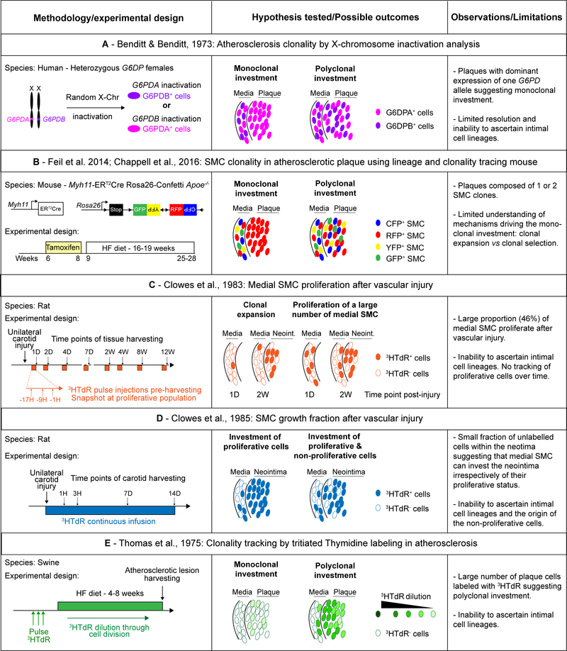
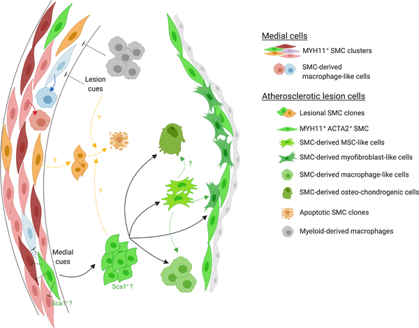
Vascular smooth muscle cells (SMC) play a critical role in controlling blood pressure and blood distribution, as well as maintaining the structural integrity of the blood vessel. SMC also participate in physiological and pathological vascular remodeling due to their remarkable ability to dynamically modulate their phenotype. During the past decade, the development of in vivo fate mapping systems for unbiased identification and tracking of SMC and their progeny has led to major discoveries as well as the reevaluation of well-established concepts about the contribution of vascular SMC in major vascular diseases including atherosclerosis. Lineage tracing studies revealed that SMC undergoes multiple phenotypic transitions characterized by the expression of markers of alternative cell types (eg, macrophage-like and mesenchymal-stem cell-like) and populate injured or diseased vessels by oligoclonal expansion of a limited number of medial SMC. With the development of high-throughput transcriptomics and single-cell RNA sequencing (scRNAseq), the field is moving forward towards in-depth SMC phenotypic characterization. Herein, we review the major observations put forth by lineage and clonality tracing studies and the evidence in support for SMC phenotypic diversity in healthy and diseased vascular tissue. We will also discuss the opportunities and remaining challenges of combining lineage tracing and single-cell transcriptomics technologies, as well as studying the functional relevance of SMC phenotypic transitions and identifying the mechanisms controlling them.

Keywords: atherosclerosis; blood pressure; cell division; cell plasticity; vascular remodeling.


Figures

Figure 1:
Figure 1:. SMC clonal expansion vs SMC proliferative capacities: a review of experimental design and possible outcomes.
This table summarizes the methodology and experimental design of seminal studies investigating: A. SMC clonality in human atherosclerotic lesions by X-chromosome (Chr) inactivation; B. SMC clonality in experimental atherosclerosis in SMC-lineage tracing mice; C. Medial SMC proliferative capacity after vascular injury by tritiated thymidine incorporation (3HTdR); D. Medial and intimal SMC growth fraction after vascular injury; and E. SMC proliferative profile within atherosclerotic lesion after tritiated thymidine pulse delivery. The table includes representations of the experimental designs and possible outcomes, as well as a summary of the main observations and limitations of these studies.
Figure 2:
Figure 2:. SMC phenotypic diversity within the media and atherosclerotic lesion.
This schematic summarizes the main finding with respect to SMC diversity. scRNAseq studies revealed that the media is composed of multiple clusters of SMC. While it is unclear whether a particular cluster of SMC has the ability to clonally expand, there is clear evidence that SMC within the atherosclerotic lesion originate from a very limited number of clones. The same SMC clone can give rise to SMC-derived fibrous cap cells, macrophage-like cells, mesenchymal-stem cell like-cell and osteochondrogenic cells. A current hypothesis is that a transition to a mesenchymal-stem cell-like cell state would precede and be required for the transition to other lesion phenotypes (green dashed arrows). In addition, it has been postulated that SMC-derived lesion cells could originate from the medial Sca1+ SMC population. This hypothesis is indicated in the schematic by the annotation of the SMC clone expanding within the lesion with “Sca1+?”. Importantly, phenotypic modulation also occurs within the media, independently of clonal expansion during atherosclerosis (red arrows). Although the mechanisms controlling the investment of atherosclerotic lesions by a very limited number of SMC clones have not been fully characterized, there is evidence that myeloid-derived plaque macrophages inhibits SMC polyclonality within atherosclerotic plaque (gray dashed arrows). It is also postulated that medial or lesion environmental cues could inhibit the proliferation and investment of multiple medial SMC within the lesion (black dashed arrows). Finally, it is unclear whether a process of clonal selection consisting in the survival of dominant clones could as well take place (yellow dashed arrows). Figure created with Biorender.

References

    1. Owens GK. Regulation of differentiation of vascular smooth muscle cells. Physiol Rev. 1995;75:487–517. - PubMed
    1. Campbell JH and Campbell GR. Smooth muscle phenotypic modulation--a personal experience. Arterioscler Thromb Vasc Biol. 2012;32:1784–9. - PubMed
    1. Chamley-Campbell J, Campbell GR and Ross R. The smooth muscle cell in culture. Physiol Rev. 1979;59:1–61. - PubMed
    1. Gomez D and Owens GK. Smooth muscle cell phenotypic switching in atherosclerosis. Cardiovasc Res. 2012;95:156–64. - PMC - PubMed
    1. Regan CP, Adam PJ, Madsen CS and Owens GK. Molecular mechanisms of decreased smooth muscle differentiation marker expression after vascular injury. J Clin Invest. 2000;106:1139–1147. - PMC - PubMed

Publication types