. 2019 Sep;39(9):1817-1830.
doi: 10.1161/ATVBAHA.119.312848. Epub 2019 Jul 25.

Formation and Resolution of Pial Microvascular Thrombosis in a Mouse Model of Thrombotic Thrombocytopenic Purpura

Affiliations

Formation and Resolution of Pial Microvascular Thrombosis in a Mouse Model of Thrombotic Thrombocytopenic Purpura

Reheman Adili et al. Arterioscler Thromb Vasc Biol. 2019 Sep.

Abstract

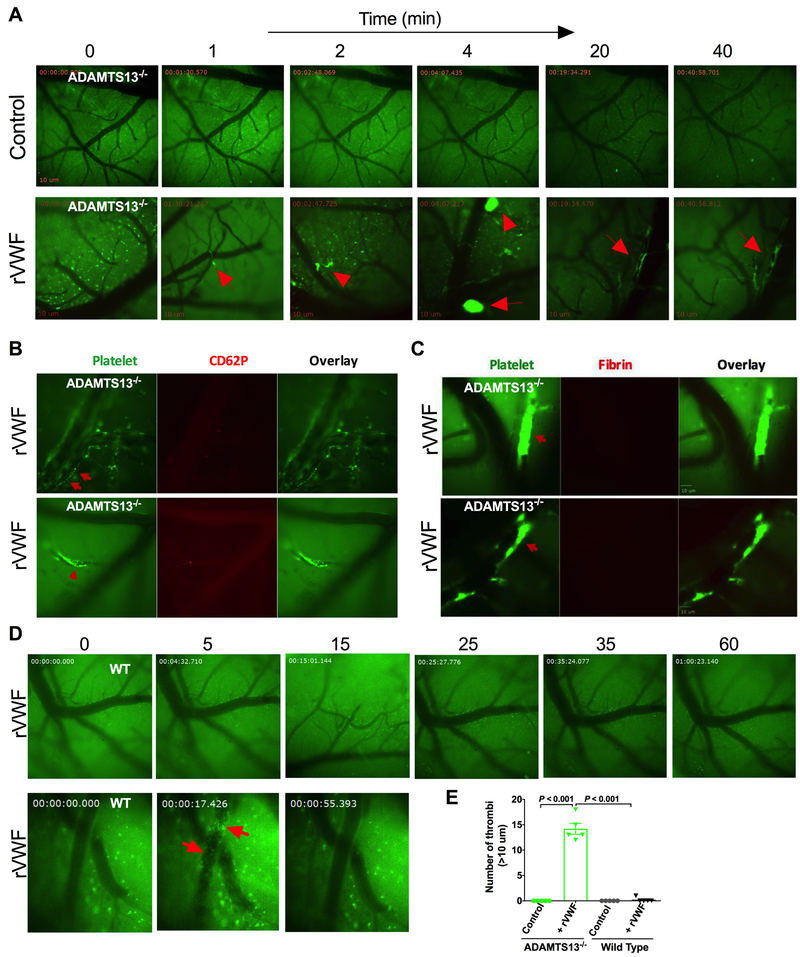
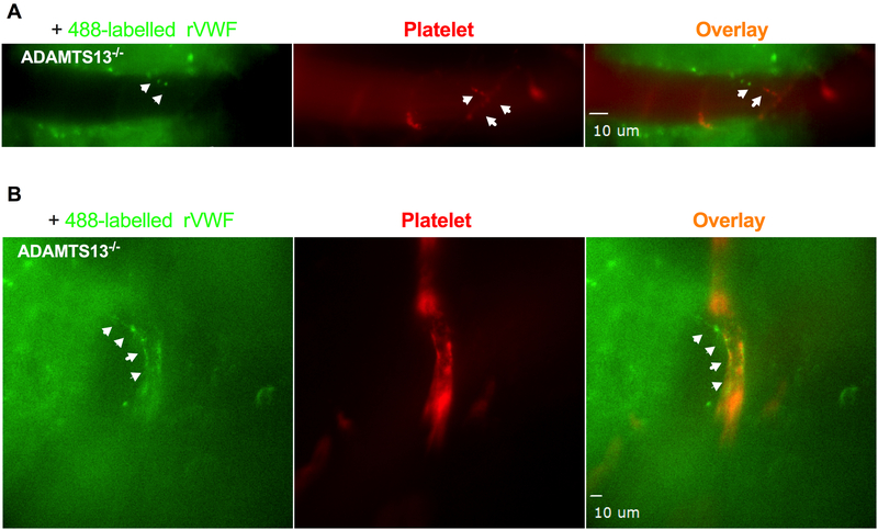
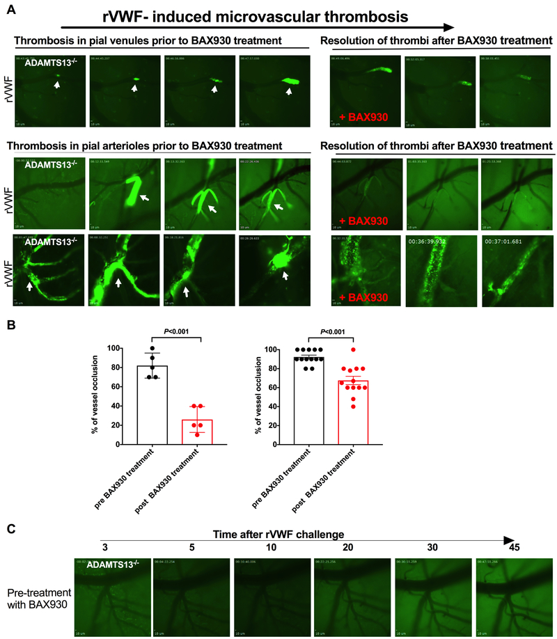
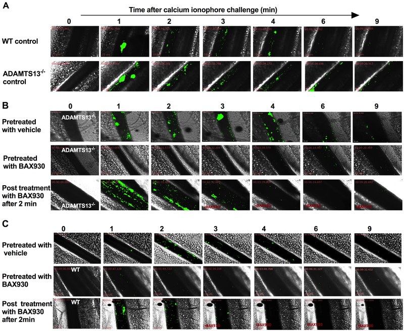
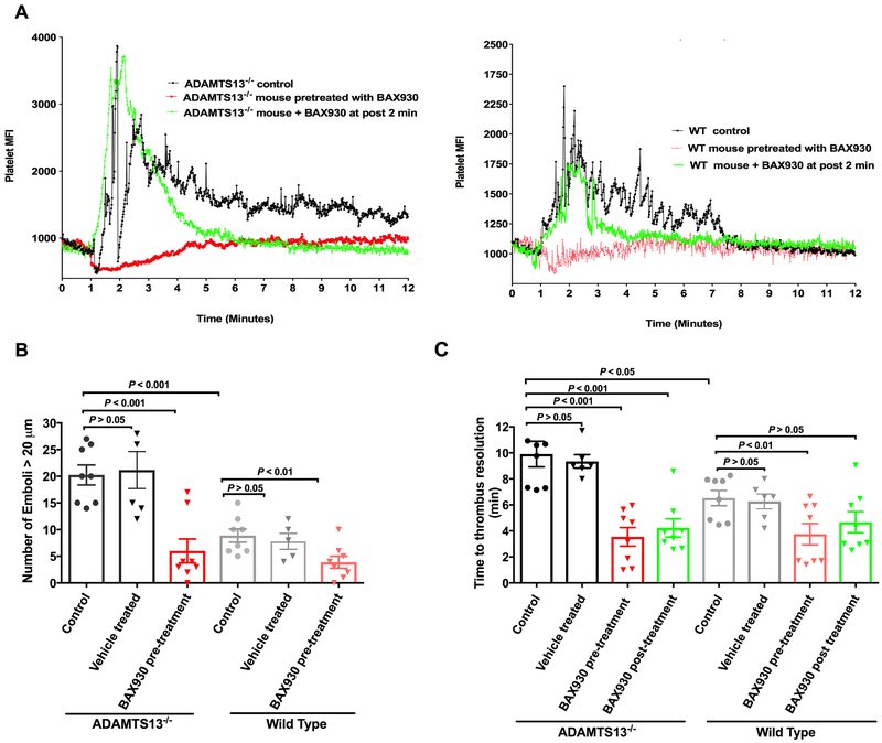
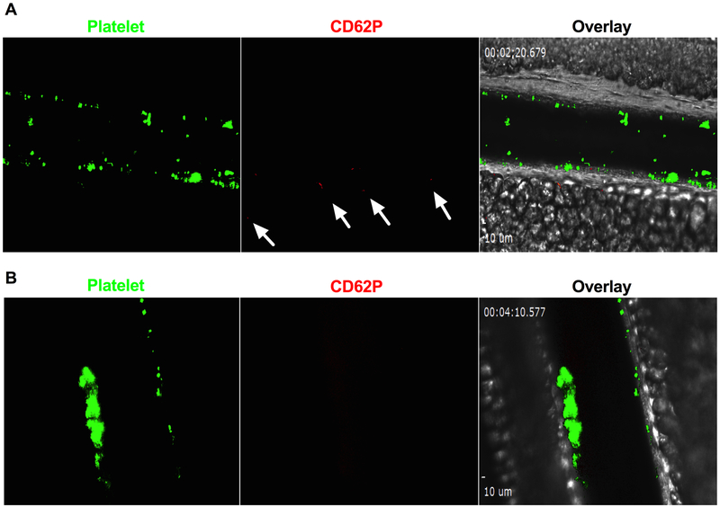
Objective: Microvascular thrombosis is the hallmark pathology of thrombotic thrombocytopenic purpura (TTP), a rare life-threatening disease. Neurological dysfunction is present in over 90% of patients with TTP, and TTP can cause long-lasting neurological damage or death. However, the pathophysiology of microvascular thrombosis in the brain is not well studied to date. Here, we investigate the formation and resolution of thrombosis in pial microvessels. Approach and Results: Using a cranial intravital microscopy in well-established mouse models of congenital TTP induced by infusion of recombinant VWF (von Willebrand factor), we found that soluble VWF, at high concentration, adheres to the endothelium of the vessel wall, self-associates, and initiates platelet adhesion resulting in the formation of pial microvascular thrombosis in ADAMTS13-/- (a disintegrin and metalloproteinase with a thrombospondin type 1 motif, member 13) mice. Importantly, VWF-mediated pial microvascular thrombosis occurred without vascular injury to the brain, and thrombi consisted of resting platelets adhered onto ultra-large VWF without fibrin in the brain in rVWF (recombinant VWF) challenged ADAMTS13-/- mice. Prophylactic treatment with recombinant ADAMTS13 (BAX930) effectively prevented the onset of the VWF-mediated microvascular thrombosis and therapeutic treatment with BAX930 acutely resolved the preexisting or growing thrombi in the brain of ADAMTS13-/- mice after rVWF challenge. The absence of platelet activation and fibrin formation within VWF-mediated thrombi and efficacy of BAX930 was confirmed with an endothelial-driven VWF-mediated microvascular thrombosis model in mice.

Conclusions: Our results provide important insight into the initiation and development of microvascular thrombi in mouse models that mimics TTP and indicate that rADAMTS13 could be an effective interventional therapy for microvascular thrombosis, the hallmark pathology in TTP.

Keywords: blood platelets; fibrin; platelet aggregation; thrombosis; von Willebrand factor.


Conflict of interest statement

Disclosure of conflict of interest

Drs. Holinstat and Adili received financial support from Shire for the research presented here. Dr. Holinstat is a consultant and equity holder for Veralox Therapeutics, although no work presented here is associated with the relationship with Veralox Therapeutics. The authors declare no additional financial interests for the work presented here. The authors were solely responsible for the content and decision to publish.

Figures

Figure 1.
Figure 1.. Initiation and development of VWF-triggered microvascular thrombosis in the brain in mouse model TTP.
A: Formation of microvascular thrombosis induced by rVWF: ADAMTS13−/− mice were intravenously injected with vehicle control or rVWF (2000 U/kg) and pial microvasculature in the brains was monitored in real-time via a cranial window under intravital microscopy for 60 minutes. Platelets were fluorescently labelled as green and pial microvessels in the brain are shown in dark. Transfusion of high dose of VWF in ADAMTS13−/− resulted in platelet adhesion, initiated thrombus formation and caused vessel occlusion in pial microvasculature in the brain under the shear condition. Thrombi formation (shown in bright green) in pail microvasculature in ADAMTS13−/− mice following the rVWF challenge are indicated by arrows. Still images were taken at the indicated times shown above after the infusion of rVWF in ADAMTS13−/− mice. B: VWF self-association on vascular wall endothelium surface and platelet P-selectin expression: Upper panel: Shortly after rVWF infusion into ADAMTS13−/− mice, soluble VWF in plasma bound to vascular wall endothelium surface and self-associated under shear to trigger platelet adhesion and thrombosis. Fluorescently-labeled platelets adhered onto the endothelial bound VWF fibers and formed long platelet strings (upper panel, indicated by arrow) that led to microvascular thrombosis (lower panel, indicated by arrow) under shear in pial microvasculature in the brains of ADAMTS13−/− mice detected with higher objective (25X) under multi-channel intravital microscopy. The majority of adhered platelets and platelets within the growing thrombi were negative for P-selectin expression on their surface except for a few single platelets shown positive for P-selectin detected by anti-mouse CD62P antibody in vivo. C: Detection of fibrin formation in microthrombi in the brain: unlike injury-induced thrombosis, fibrin formation was not detected in rVWF-triggered microvascular thrombosis in pial microvessels in the brain. Fibrin formation within platelet thrombi in pial microvasculature in three different ADAMTS13−/− mice was examined in vivo using fluorescently labeled anti-mouse fibrin antibody, and no fibrin formation was detected within thrombi (n= 5) in the period of 60 min recording. D: Platelet adhesion and thrombosis was not detected in the brain microvessels in wild type (WT) mice after rVWF (2000 U/kg) challenge (upper panel). Bolus injection of rVWF resulted in an immediate presence of circulating platelet micro aggregates in pial microvasculature (lower panel) detected with higher objective (25X) but quickly cleared from blood circulation in less than a minute. rVWF challenge in WT mice did not result in VWF fibrillar and platelet string formation on endothelial surface on vessel wall in vivo. E: Quantitative analysis of pial microvascular thrombi larger than 10 μm in diameter following the vehicle control or rVWF challenge in ADAMTS13−/− and WT mice.
Figure 2.
Figure 2.. Exogenous recombinant VWF bind to the endothelial surface of pial microvascular wall and VWF self-associate to trigger platelet adhesion and thrombosis.
Alexa Fluor 488-conjugated rVWF (Green) were intravenously injected to ADAMTS13−/− mice and monitored in real-time by intravital microscopy for endothelial interaction, platelet adhesion and thrombosis in pial microvascular circulation. Platelets in ADAMTS13−/− mice were fluorescently labeled using a DyLight 649-conjugated GP1bβ antibody (Red). A: Fluorescently labeled rVWF bound to vascular wall endothelium surface, self-associated to form ULVWF fibers (left panel, indicated by arrows). Adhered platelets on endothelial-bound ULVWF are shown in red (middle panel, indicated by arrows). Adhered platelets on ULVWF are shown as platelet string in overlaid image. (right panel indicated by arrows). B: VWF fibers along the vessel wall within microvascular thrombosis detected with higher objective (25X) under multi-channel intravital microscopy.
Figure 3.
Figure 3.. Recombinant ADAMTS13 treatment effectively resolves pre-existing and growing microvascular thrombi in the brain in a mouse model of TTP.
A: Representative images of microvascular thrombosis formed in the brain of 3 different ADAMTS13−/− mice following the transfusion of rVWF and the effect of BAX930 treatment. Pial microvasculature in the brain in ADAMTS13−/− mice is shown in dark and formed thrombi are shown in bright green. Thrombus formation in pial microvessels prior to BAX930 treatment are shown on the left and effect of rADAMTS13 treatment on thrombosis are shown on the right. Thrombi were observed for up to 60 minutes under the cranial window intravital microscopy. Upper panel: Thrombus formation in the pial venule was monitored from onset until it reached more than 70% occlusion, at which point an immediate tail vein injection of BAX930 was given. Thrombus growth was immediately attenuated, and the thrombi resolved. Lower panel arteriole 1: Thrombus formation was captured occluding the pial arteriole at the bifurcation. The thrombus was visibly loose, forming small emboli but consistently growing for the next 20 min before BAX930 treatment. BAX930 treatment resolved the embolic thrombi, improved the blood flow, and decreased the size of the thrombi over time. Arteriole 2: Vessel occlusion by thrombus in pial arteriole was captured in ADAMTS13−/− mice pretreated with the vehicle control. BAX930 treatment was initiated after vessel occlusion to observe the resolution of the thrombus. The thrombus was resolved, and blood flow was partially restored by BAX930 treatment. Pial microvasculature in the brain in ADAMTS13−/− mice is shown in dark and time elapsed since the VWF infusion is indicated. B: Quantitative analysis of the effect of BAX930 treatment on vessel occlusion. Left: comparison of percentage of vessel (<10 μm in diameter) occlusion by thrombi pre and post BAX930 treatment. (P< 0.001 n= 5;) Right: comparison of percentage of vessel (>10 μm in diameter) occlusion by thrombi pre and post BAX930 treatment. (P< 0.001 n= 13;) C: Prophylactic treatment with BAX930 protected ADAMTS13−/− mice from microvascular thrombosis in the brain following the VWF challenge. Times after rVWF challenge in ADAMTS13−/− mice is indicated above. 5 mice in each group were studied.
Figure 4.
Figure 4.. Recombinant ADAMTS13 (a disintegrin and metalloproteinase with a thrombospondin type 1 motif, member 13) treatment attenuates platelet accumulation on newly released endothelial-bound ULVWF (ultra-large von Willebrand factor) in vivo.
Mesenteric venules of ADAMTS13−/− mice were topically treated with calcium ionophore to release ULVWF from the endothelium of vessel wall and platelet adhesion and accumulation on endothelial-bound ULVWF was monitored in real-time under intravital microscopy. Florescent platelets are shown in green and sequential images taken at the indicated times after the application of calcium ionophore are aligned. A: Representative sequential images of VWF mediated platelet adhesion and thrombosis in WT (upper panel) and ADAMTS13−/− mice (lower panel). Ionophore-provoked thrombosis was enhanced and prolonged in ADAMTS13 deficiency. B: Representative sequential images of ionophore-provoked thrombosis in ADAMTS13−/− mice treated with vehicle control or BAX930 treatment. Upper panel: ADAMTS13−/− mice pretreated with vehicle control; Middle panel: ADAMTS13−/− mice pretreated with BAX930 10 min prior to calcium ionophore application; Lower panel: ADAMTS13−/− mice treated with BAX930 2 min after the onset of thrombosis following the calcium ionophore application. Prophylactic and therapeutic BAX930 treatment in ADAMTS13−/− mice effectively prevented and immediately resolved the growing thrombi in mesenteric venules. C: Representative sequential images of ionophore-provoked thrombosis in WT mice treated with vehicle control or BAX930 treatment. Upper panel: WT mice pretreated with vehicle control; Middle panel: WT mice pretreated with BAX930 10 min prior to calcium ionophore application; Lower panel: WT mice treated with BAX930 2 min after the onset of thrombosis following the calcium ionophore application. Prophylactic BAX930 treatment in WT effectively inhibited thrombosis in response to calcium ionophore. Therapeutic BAX930 treatment in WT mice immediately resolved the growing thrombi in mesenteric venules. 8 mice were examined in each group.
Figure 5.
Figure 5.. Quantitative analysis of platelet accumulation and emboli formation in ionophore-provoked ULVWF-mediated mesenteric microvascular thrombosis model:
A: Representative traces of dynamics of platelet accumulation quantified by platelet fluorescent intensity over time in vehicle treated control ADAMTS13−/− mice (Left) and WT mice (Right) are shown in black color. Topical application of calcium ionophore on the mesenteric venules resulted in a sharp increase in platelet mean fluorescence intensity (MFI) as a result of platelet thrombus formation followed by drop in platelet MFI by many spikes representing embolization of thrombi in the vessel segment being monitored until ultimately returning to baseline level. The peak platelet MFI in ADAMTS13−/− mice was significantly higher than that of WT mice (P<0.05) due to enhanced thrombotic response in ADAMTS13 deficiency. Fluctuation in platelet MFI was more notable in ADAMTS13−/− mice (as compared to WT mice) due to larger size and prolonged emboli formation. Prophylactic BAX930 treatment in both ADAMTS13−/− mice and WT mice (shown in red) effectively prevented platelet adhesion and formation of platelet thrombi (ADAMTS13−/− P<0.001, WT P<0.01). Therapeutic administration of BAX930 in both ADAMTS13−/− mice and WT (shown in green) at 2 min after the onset of thrombosis induced by ionophore application acutely attenuated thrombus formation and accelerated thrombus resolution. B: Quantification of emboli formation. Number of large emboli (>20 μm in diameter) formed in mesenteric venules of ADAMTS13−/− (column 1 to 3) or WT mice (column 4 to 6) that untreated or pretreated with vehicle control (black: ADAMTS13−/−, grey: WT) or pretreated with BAX930 (shown in red) in response to calcium ionophore treatment. Significantly more emboli were formed in ADAMTS13−/− mice when compared to WT. Prophylactic BAX930 treatment effectively inhibited thrombus formation in both ADAMTS13−/− and WT mice. C. Thrombus resolution time. Time required for the platelet the fluorescence returning to near baseline following the topical application in ADAMTS13−/− (column 1 to 4) or WT mice (column 5 to 8) that untreated or pretreated with vehicle control (black: ADAMTS13−/−, grey: WT) or pretreated with BAX930 (shown in red) or BAX930 post treatment (green). Time for thrombus resolution was significantly longer in ADAMTS13−/− mice when compared to WT. Thrombus resolution time was significantly shorter in both pre and post BAX930 treated ADAMTS13−/− mice. Thrombus resolution time in WT was significantly shortened by prophylactic BAX930 treatment.
Figure 6:
Figure 6:. Platelet surface P-selectin expression and fibrin formation in ionophore-provoked microvascular thrombosis model.
A: Platelets in ADAMTS13−/− were fluorescently labelled as green and platelet surface P-selectin was detected by anti-P-selectin antibody (red) in vivo. P-selectin expression was not detected on the majority of platelets within thrombi except for the few platelets shown to be positive for P-selectin (indicated by arrows). B: Fibrin formation in thrombi: Fibrin formation was not detected in ionophore-provoked ULVWF-mediated microvascular thrombosis in mesenteric microvascular thrombosis in ADAMTS13−/− mice (n=3). Positive images of P-selectin and fibrin formation were confirmed by laser injury induced thrombosis in the same mice (shown in the supplementary).

Similar articles

Cited by

References

    1. Tsai HM. Current concepts in thrombotic thrombocytopenic purpura. Annu Rev Med. 2006;57:419–436 - PMC - PubMed
    1. Sadler JE, Moake JL, Miyata T, George JN. Recent advances in thrombotic thrombocytopenic purpura. Hematology Am Soc Hematol Educ Program. 2004:407–423 - PubMed
    1. Kremer Hovinga JA, Coppo P, Lammle B, Moake JL, Miyata T, Vanhoorelbeke K. Thrombotic thrombocytopenic purpura. Nat Rev Dis Primers. 2017;3:17020. - PubMed
    1. Levy GG, Nichols WC, Lian EC, Foroud T, McClintick JN, McGee BM, Yang AY, Siemieniak DR, Stark KR, Gruppo R, Sarode R, Shurin SB, Chandrasekaran V, Stabler SP, Sabio H, Bouhassira EE, Upshaw JD Jr., Ginsburg D, Tsai HM. Mutations in a member of the adamts gene family cause thrombotic thrombocytopenic purpura. Nature. 2001;413:488–494 - PubMed
    1. Zheng X, Chung D, Takayama TK, Majerus EM, Sadler JE, Fujikawa K. Structure of von willebrand factor-cleaving protease (adamts13), a metalloprotease involved in thrombotic thrombocytopenic purpura. J Biol Chem. 2001;276:41059–41063 - PubMed

Publication types