

Effect of female sex hormones on the developmental cycle of Chlamydia abortus compared to a penicillin-induced model of persistent infection
- PMID: 31340824
- PMCID: PMC6657046
- DOI: 10.1186/s12917-019-2013-7
Effect of female sex hormones on the developmental cycle of Chlamydia abortus compared to a penicillin-induced model of persistent infection
Abstract
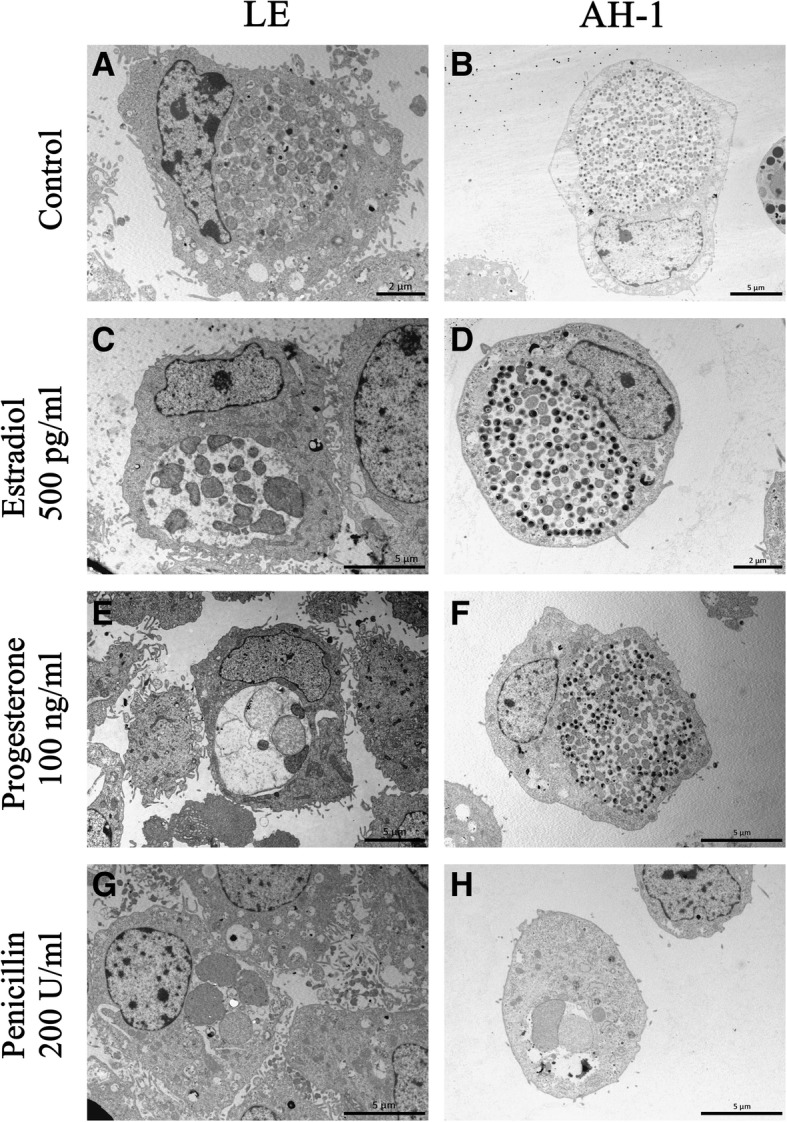
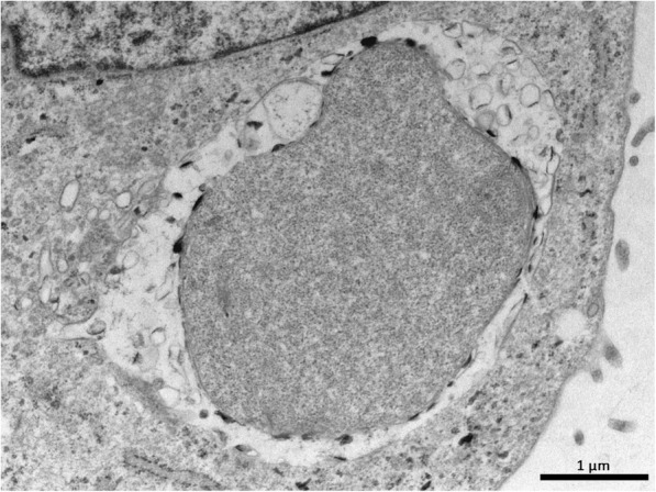
Background: Chlamydia abortus, an obligate intracellular pathogen with an affinity for placenta, causes reproductive failure. In non-pregnant animals, an initial latent infection is established until the next gestation, when the microorganism is reactivated, causing abortion. The precise mechanisms that trigger the awakening of C. abortus are still unknown. Sexual hormones such as estradiol and progesterone have been shown to affect the outcome of infection in other species of the family Chlamydiaceae, while estrogens increase chlamydial infection, progesterone has the opposite effect. To try to establish whether there is a relationship between these events and the latency/ reactivation of C. abortus in the reproductive tract of small ruminants, ovine endometrial (LE) and trophoblastic (AH-1) cells were treated with estradiol or progesterone prior to their infection with C. abortus. The results are compared with those obtained for treatment with penicillin prior to infection, which is a well-established model for studying persistent infection in other chlamydial species. Cells were examined by transmission electron microscopy, and an mRNA expression analysis of 16 genes related to the chlamydial developmental cycle was made.
Results: The changes observed in this study by the action of sex hormones seem to depend on the type of cell where the infection develops. In addition, while the changes are morphologically similar to those induced by treatment with penicillin, the patterns of gene expression are different. Gene expression patterns therefore, seem to depend on the persistence induced models of C. abortus used. Hormone treatments induced aberrant forms in infected endometrial cells but did not affect the chlamydial morphology in trophoblast cells. At the genetic level, hormones did not induce significant changes in the expression of the studied genes.
Conclusions: The results suggest that penicillin induces a state of persistence in in vitro cultured C. abortus with characteristic morphological features and gene transcriptional patterns. However, the influence of hormones on the C. abortus developmental cycle is mediated by changes in the host cell environment. Furthermore, a persistent state in C. abortus cannot be characterised by a single profile of gene expression pattern, but may change depending on the model used to induce persistence.
Keywords: Chlamydia abortus; Female sex hormones; Ovine enzootic abortion; Penicillin; Persistence.
Conflict of interest statement
The authors declare that they have no competing interests.
Figures
References
-
- Santoro M, Laccarino D, Di Nocera F, Degli Uberti B, Lucibelli MG, Borriello G, De Luca G, D’Amore M, Cerrone A, Galiero G. Molecular detection of Chlamydia abortus in a stranded Mediterranean striped dolphi Stenella coeruleoalba. Dis Aquat Org. 2019;132:203–208. doi: 10.3354/dao03320. - DOI - PubMed
Publication types
MeSH terms
Substances
Grants and funding
LinkOut - more resources
Full Text Sources
