. 2019 Sep 13;294(37):13545-13559.
doi: 10.1074/jbc.RA119.009725. Epub 2019 Jul 24.

The crystal structure of the protein kinase HIPK2 reveals a unique architecture of its CMGC-insert region

Affiliations

The crystal structure of the protein kinase HIPK2 reveals a unique architecture of its CMGC-insert region

Christopher Agnew et al. J Biol Chem. .

Abstract

The homeodomain-interacting protein kinase (HIPK) family is comprised of four nuclear protein kinases, HIPK1-4. HIPK proteins phosphorylate a diverse range of transcription factors involved in cell proliferation, differentiation, and apoptosis. HIPK2, thus far the best-characterized member of this largely understudied family of protein kinases, plays a role in the activation of p53 in response to DNA damage. Despite this tumor-suppressor function, HIPK2 is also found overexpressed in several cancers, and its hyperactivation causes chronic fibrosis. There are currently no structures of HIPK2 or of any other HIPK kinase. Here, we report the crystal structure of HIPK2's kinase domain bound to CX-4945, a casein kinase 2α (CK2α) inhibitor currently in clinical trials against several cancers. The structure, determined at 2.2 Å resolution, revealed that CX-4945 engages the HIPK2 active site in a hybrid binding mode between that seen in structures of CK2α and Pim1 kinases. The HIPK2 kinase domain crystallized in the active conformation, which was stabilized by phosphorylation of the activation loop. We noted that the overall kinase domain fold of HIPK2 closely resembles that of evolutionarily related dual-specificity tyrosine-regulated kinases (DYRKs). Most significant structural differences between HIPK2 and DYRKs included an absence of the regulatory N-terminal domain and a unique conformation of the CMGC-insert region and of a newly defined insert segment in the αC-β4 loop. This first crystal structure of HIPK2 paves the way for characterizing the understudied members of the HIPK family and for developing HIPK2-directed therapies for managing cancer and fibrosis.

Keywords: CMGC superfamily; DYRK1A; cancer; fibrosis; homeodomain-interacting protein kinase 2 (HIPK2); inhibitor; neurodegenerative disease; serine/threonine protein kinase; structural biology.


Conflict of interest statement

The authors declare that they have no conflicts of interest with the contents of this article. The content is solely the responsibility of the authors and does not necessarily represent the official views of the National Institutes of Health.

Figures

Figure 1.
Figure 1.
The HIPKs are members of the CMGC kinase family. A, phylogenetic tree of the CMGC kinase family branch that includes the HIPK family. B, domain architecture of the four HIPK family members. The construct boundaries for crystallized HIPK2 are indicated.
Figure 2.
Figure 2.
Structure of HIPK2 kinase domain. A, crystal structure of HIPK2 kinase domain bound to inhibitor CX-4945 in the active site. The helix C (αC) is shown in blue, the activation loop in red, the CMGC-insert in violet, and the inhibitor in yellow. B, active site of the HIPK2 kinase showcasing the bound CX-4945 inhibitor. Key residues are depicted as sticks and numbered. Hydrogen bonds are shown as dashed black lines. C, sequence alignment of active-site residues for selected CMGC kinases. D, a hydrogen bond network in HIPK2 that stabilizes the activation loop conformation via an interaction between the CMGC arginine (Arg368) and the phosphorylated activation loop tyrosine (Tyr361).
Figure 3.
Figure 3.
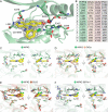
Binding of CX-4945 to HIPK2 ATP-pocket. A, zoomed-in view of the active site in the crystal structure of the HIPK2 kinase in complex with CX-4945. Inhibitor is shown with the 2FoFc map contoured to 1.5σ. Hydrogen bonds are shown as black dashes, and a water molecule is shown as a red sphere. B, for clarity, identifiers of all residues in structure images shown in C–F were reduced to single letters. A table summarizing the correspondence of the letters to individual residues in each structure is shown here. C–F, zoomed-in views of the active sites of the indicated CMGC kinases crystallized in complex with the CX-4945 inhibitor. Inhibitor binding is shown face-on and upon 70° rotation. C, HIPK2 bound to CX-4945; D, CK2α bound to CX-4945 (PDB code 3NGA); E, CLK1 bound to CX-4945 (PDB code 6FYV); F, Pim1 bound to CX-4945 (PDB code 5O11).
Figure 4.
Figure 4.
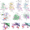
HIPK2 contains a unique CMGC-insert. A, side-by-side comparison of the crystal structure of the CX-4945–bound HIPK2 kinase domain with DYRK1A (PDB code 4MQ1), DYRK2 (PDB code 3K2L), and DYRK3 (PDB code 5Y86). B, comparison of active site between HIPK2 and DYRK2; numbering in parentheses corresponds to DYRK2. C, zoomed-in view of the secondary structural elements in the CMGC-insert visualized in the CX-4945–bound HIPK2 kinase structure and its trajectory on the kinase C-lobe. D, comparison between the CMGC-insert regions in HIPK2 and DYRK2 kinases made by overlay of the CX-4945–bound HIPK2 kinase structure and the DYRK2 structure (PDB code 3K2L). E, phosphorylation of CMGC-insert serine residues observed in the structures of the CX-4945–bound HIPK2 kinase, DYRK2 (PDB code 3K2L), and DYRK3 (PDB code 5Y86).
Figure 5.
Figure 5.
Unique structural features of the HIPK family. A, sequence alignment of a region within the C-lobe in HIPK sequences from diverse phyla, as revealed by our analysis of evolutionary constraints acting on HIPK sequences (Fig. S6). Three sets of related sequences are shown in the hierarchical alignment: (i) a foreground set of 1498 HIPK sequences that share a co-conserved pattern, as defined by the Bayesian pattern-partitioning procedure, (ii) a background set of 14,296 CMGC sequences, and (iii) a display set of HIPK homologs from diverse phyla. Only the display sequences are explicitly shown in the alignment. The foreground (HIPK) and background (CMGC) alignments are shown as residue frequencies below the display alignment. Residue frequencies are indicated in integer tenths where, for example, a 9 indicates 90–100% occurrence of the corresponding residue, at the corresponding position, in weighted foreground or background sequences. Patterns identified as unique to HIPK sequence are indicated by black dots above the alignment and highlighted in the display alignment. Evolutionary constraints on the pattern residues are displayed as red histograms above the alignment, where the height of the histogram indicates the strength of HIPK-specific constraints at the corresponding position. B, evolutionarily constrained residues in the HIPK2 kinase C-lobe that form specific interactions with the CMGC-insert (shown in surface mode) are shown as sticks. C, distinguishing residues in the αC–β4 loop in HIPK kinases define a unique insert region. Insert sequences are indicated in lowercase letters, and their frequency is not scored due to their absence in all non-HIPK kinase sequences. D, the residues stabilizing the unique conformation of the αC–β4 loop insert in HIPK2 are shown as sticks. E, comparison of the αC–β4 loop structure in the HIPK2 kinase with DYRK1A (PDB code 3ANQ) and GSK3β (PDB code 1GNG).

References

    1. Manning G., Whyte D. B., Martinez R., Hunter T., and Sudarsanam S. (2002) The protein kinase complement of the human genome. Science 298, 1912–1934 10.1126/science.1075762 - DOI - PubMed
    1. Kim Y. H., Choi C. Y., Lee S. J., Conti M. A., and Kim Y. (1998) Homeodomain-interacting protein kinases, a novel family of co-repressors for homeodomain transcription factors. J. Biol. Chem. 273, 25875–25879 10.1074/jbc.273.40.25875 - DOI - PubMed
    1. Blaquiere J. A., and Verheyen E. M. (2017) Homeodomain-interacting protein kinases: diverse and complex roles in development and disease. Curr. Top. Dev. Biol. 123, 73–103 10.1016/bs.ctdb.2016.10.002 - DOI - PubMed
    1. Hofmann T. G., Glas C., and Bitomsky N. (2013) HIPK2: A tumour suppressor that controls DNA damage-induced cell fate and cytokinesis. Bioessays 35, 55–64 10.1002/bies.201200060 - DOI - PubMed
    1. Shang Y., Doan C. N., Arnold T. D., Lee S., Tang A. A., Reichardt L. F., and Huang E. J. (2013) Transcriptional corepressors HIPK1 and HIPK2 control angiogenesis via TGF-β-TAK1-dependent mechanism. PLoS Biol. 11, e1001527 10.1371/journal.pbio.1001527 - DOI - PMC - PubMed

Publication types

MeSH terms