Review
. 2019 Jul 24;11(502):eaau5485.
doi: 10.1126/scitranslmed.aau5485.

The 1918 influenza pandemic: 100 years of questions answered and unanswered

Affiliations
Review

The 1918 influenza pandemic: 100 years of questions answered and unanswered

Jeffery K Taubenberger et al. Sci Transl Med. .

Abstract

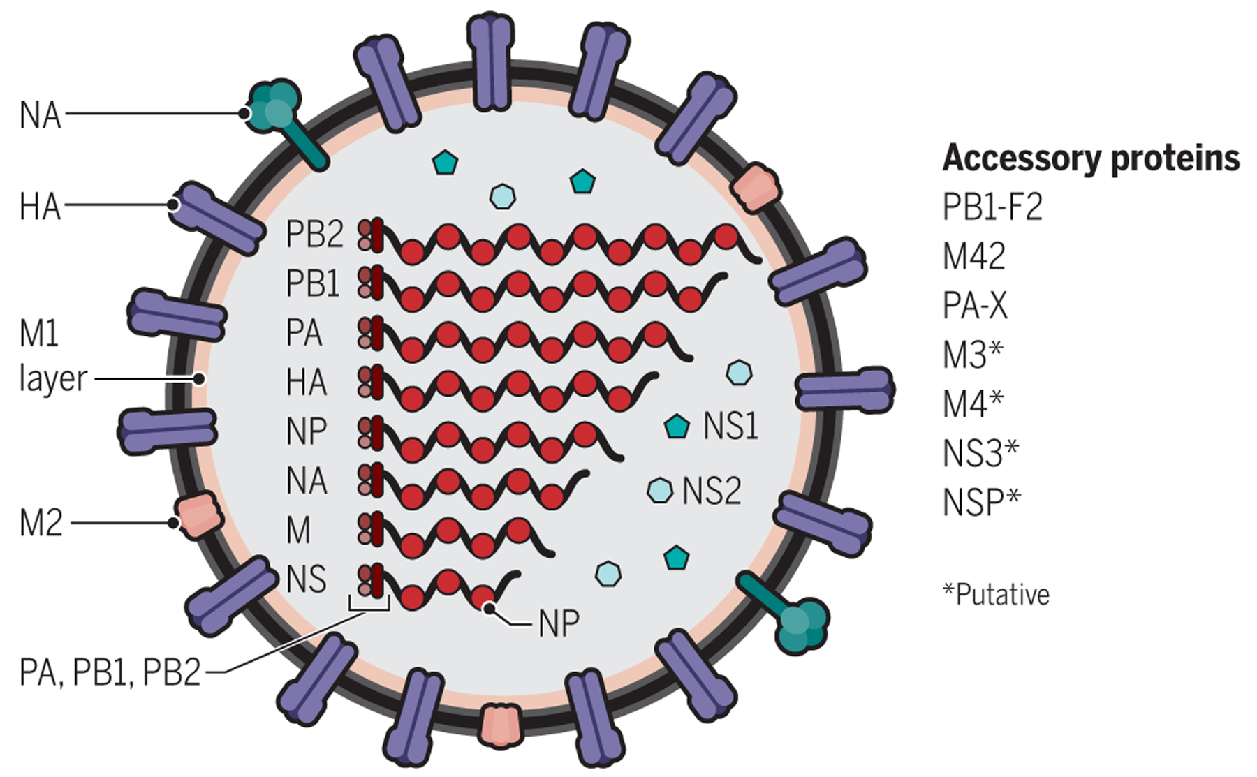
The 2018-2019 period marks the centennial of the "Spanish" influenza pandemic, which caused at least 50 million deaths worldwide. The unprecedented nature of the pandemic's sudden appearance and high fatality rate serve as a stark reminder of the threat influenza poses. Unusual features of the 1918-1919 pandemic, including age-specific mortality and the high frequency of severe pneumonias, are still not fully understood. Sequencing and reconstruction of the 1918 virus has allowed scientists to answer many questions about its origin and pathogenicity, although many questions remain. This Review summarizes key findings and still-to-be answered questions about this deadliest of human events.


Figures

Fig. 1.
Fig. 1.. Structure of influenza A viruses.
Influenza A viruses are composed of 8 gene segments and 11 or more proteins. The surface glycoproteins hemagglutinin (HA) and neuraminidase (NA) are the major antigenic targets of the host immune response to influenza A virus infection. NA is the target of the antiviral drugs oseltamivir and zanamivir. Matrix 1 (M1) is a structural protein, and matrix 2 (M2) is an ion channel protein. Nucleoprotein (NP) encapsidates the viral RNA. Each gene segment is associated with a trimeric RNA-dependent RNA polymerase complex consisting of the PB1, PB2, and PA proteins. The nonstructural 1 (NS1) protein has pleiotropic functions, including binding to double-stranded RNA, enhancement of viral mRNA translation, inhibition of host mRNA processing, and blocking the type I interferon response of the host. NS2 [also referred to as nuclear export protein (NEP)] is found in virions and facilitates nuclear export of viral ribonucleoprotein complexes. Another small viral protein, PB1-F2, is variably encoded within the PB1 gene by an alternative reading frame; it targets the mitochondrial inner membrane and may play a role in apoptosis during influenza A viral infection. Influenza pandemics over the past century have emerged in several different ways. They can emerge directly due to influenza A viruses that inhabit wild waterfowl switching to a human host, as likely occurred in the 1918 pandemic. They can result from acquisition of gene segments through reassortment of different HA subtypes with or without reassortment-associated acquisition of other gene segments (so-called antigenic shift). They can also result from complex reassortment and host adaptation events, such as occurred in the 2009 pandemic, involving reassortment between human, swine, and avian influenza A viruses. Major HA changes in seasonal endemic viruses, arising from intrasubtypic reassortment, may also cause pandemics, as happened in 1946 (147). Since 1918, there have been pandemics caused by 1918 descended viruses: in 1957 (H2N2), in 1968 (H3N2), the reemergence of H1N1 in 1977, and the emergence of a new H1N1 virus in 2009 (12). Between pandemics, annual seasonal influenza A viruses are generated by continuous viral genetic mutations (antigenic drift) and by intrasubtypic reassortments (–150). The figure is adapted from (10).
Fig. 2.
Fig. 2.. Influenza pandemics of the past 100 years.
The 1918 “Spanish flu” pandemic was caused by a founder H1N1 influenza A virus. The three subsequent pandemics of 1957, 1968, and 2009 (black arrows) were caused by descendants of the 1918 virus, which acquired one or more genes through reassortment (12). Colored horizontal lines reflect the years of annual epidemics of seasonal influenza that occurred after each pandemic. In 1977, pre-1957 human H1N1 viruses reemerged presumably through accidental release of an older human strain from a laboratory (151). This resulted in a 20-year gap in H1N1 circulation as indicated by the discontinuous turquoise line. Consequently, human H3N2 viruses co-circulated with human 1918 lineage seasonal H1N1 viruses from 1977 to 2009, when this lineage was replaced by the new swine-origin H1N1 2009 pandemic virus. Since 2009, H3N2 viruses have co-circulated with 2009 pandemic lineage H1N1 viruses in humans.
Fig. 3.
Fig. 3.. Evolution and annual mortality of four pandemic influenza virus strains.
The four pandemic strains shown are descended from the 1918 pandemic virus and arose by antigenic shift between 1955 and 2016. (A) Antigenic changes in post-pandemic viruses are indicated. The colored bars represent the prevalence of the 1957 H2N2 pandemic virus (red), the 1968 H3N2 pandemic virus (dark blue), the unexpected return of a 1950s-era descendant of the 1918 pandemic virus, presumably released accidentally from a laboratory (turquoise), and the 2009 H1N1 pandemic virus (orange). Changes in antigenic drift of sufficient magnitude to require reformulation of the annual vaccine are indicated on the y axis. Notably, the 1968 H3N2 virus has been drifting at a greater rate (an average of 0.7 genetic changes per year) than the other three pandemic viruses (an average of 0.27 genetic changes per year for the three combined). Antigenic changes in post-pandemic influenza viruses are associated with antigenic drift, which introduces new epitopes or new glycosylation sites, and by intrasubtypic reassortment of an antigenically different HA of the same subtype, represented by vertical white lines. (B) Annual excess U.S. mortality rates attributed to influenza from 1955 to 2016. Pandemic eras since 1957 are represented by different colors, as shown by the key below the graph. Data are missing for some early years. Data were obtained from the U.S. Centers for Disease Control and Prevention and reflect excess all-cause mortality, the most common calculation method available for this time period (which may overestimate mortality). Figures are modified and updated from (152).
Fig. 4.
Fig. 4.. Pathological features of the 1918 pandemic influenza virus.
(A) Photomicrograph of a hematoxylin and eosin (H&E)–stained section of lung from autopsy material from a 1918 influenza case with acute pneumonia. The image shows a necrotizing bronchiolitis with massive infiltration of neutrophils through the wall and into the lumen of a bronchiole. Original magnification, ×200. (B) Immunohistochemical staining of influenza viral antigens in a bronchiole from lung autopsy material from a 1918 influenza case. Viral antigens stain human respiratory epithelial cells reddish brown on a hematoxylin-stained background. Original magnification, ×200. (C) H&E-stained section of lung obtained from a 1918 influenza case at autopsy. The image shows diffuse alveolar damage with acute pulmonary edema and hemorrhage filling the lung air spaces. The alveolar air spaces contain edema fluid, strands of fibrin, red blood cells, and inflammatory cells. Original magnification, ×100. (D) Immunohistochemical staining of a section of lung obtained from a 1918 influenza case showing influenza viral antigens in alveolar epithelial cells and lung alveolar macrophages. Viral antigens stain alveolar cells reddish brown on a hematoxylin-stained background. Original magnification, ×1000. (E) Section of lung obtained from a 1918 influenza case with acute pneumonia. The image shows a bacterial bronchopneumonia characterized by a necrotizing bronchitis and bronchiolitis and massive infiltration of neutrophils into the lung air spaces of surrounding alveoli. Original magnification, ×100. (F) Gram staining of a lung tissue section obtained at autopsy from a 1918 influenza case with pneumonia. Gram-positive cocci morphologically compatible with S. pyogenes are purple. Original magnification, ×1000. Figure panels represent histological findings for autopsy lung samples from 1918 fatal influenza cases.
Fig. 5.
Fig. 5.. Age-specific influenza mortality (1918–1922).
The 1918 Spanish influenza pandemic appeared in Breslau (now Wrocław), Poland, in October 1918, causing high mortality. The “W-shaped” age-specific mortality pattern indicated in the graph was seen worldwide. Influenza age-specific mortality is usually U-shaped with higher mortality in infants and the elderly. A third peak of mortality in young adults (peaking at about age 27) was uniquely associated with the 1918 pandemic. Pandemic recurrences 15 and 26 months later were, however, associated with lower overall mortality and the virtual disappearance by 1922 of mortality in young adults. Modified from (84).

References

    1. Jordan EO, Epidemic Influenza: A Survey (American Medical Association, 1927).
    1. Taubenberger JK, Morens DM, 1918 influenza: The mother of all pandemics. Emerg. Infect. Dis 12, 15–22 (2006). - PMC - PubMed
    1. Morens DM, Taubenberger JK, Fauci AS, Predominant role of bacterial pneumonia as a cause of death in pandemic influenza: Implications for pandemic influenza preparedness. J. Infect. Dis 198, 962–970 (2008). - PMC - PubMed
    1. Viboud C, Eisenstein J, Reid AH, Janczewski TA, Morens DM, Taubenberger JK, Age- and sex-specific mortality associated with the 1918–1919 influenza pandemic in Kentucky. J. Infect. Dis 207, 721–729 (2013). - PMC - PubMed
    1. Chandra S, Kuljanin G, Wray J, Mortality from the influenza pandemic of 1918–1919: The case of India. Demography 49, 857–865 (2012). - PubMed

Publication types

MeSH terms