. 2019 Jul 24;9(1):10722.
doi: 10.1038/s41598-019-47224-2.

Plasticity of patient-matched normal mammary epithelial cells is dependent on autologous adipose-derived stem cells

Affiliations

Plasticity of patient-matched normal mammary epithelial cells is dependent on autologous adipose-derived stem cells

Annika Kengelbach-Weigand et al. Sci Rep. .

Abstract

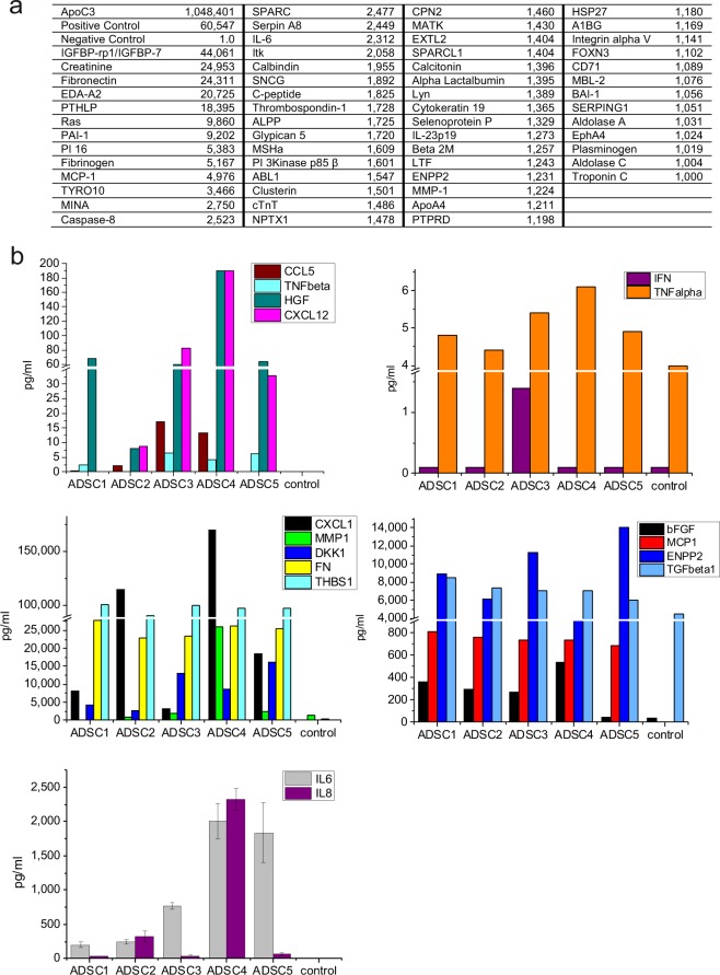
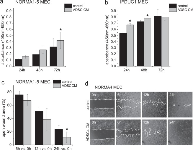
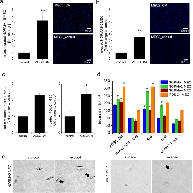
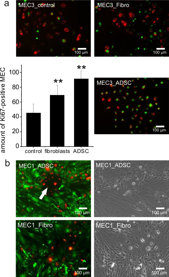
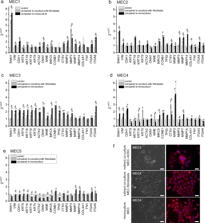
Due to the increasing clinical application of adipose-derived stem cells (ADSC), e.g. lipotransfer for breast reconstruction, this study aimed to gain novel insights regarding ADSC influence on breast tissue remodeling and determine patient-dependent factors affecting lipotransfer as well as begin to address its oncological risks. The ADSC secretome was analyzed from five normal breast reduction patients and contained elevated levels of growth factors, cytokines and proteins mediating invasion. ADSC/ADSC secretomes were tested for their influence on the function of primary mammary epithelial cells, and tumor epithelial cells using cell culture assays. ADSC/ADSC secretomes significantly stimulated proliferation, transmigration and 3D-invasion of primary normal and tumor epithelial cells. IL-6 significantly induced an EMT and invasion. The ADSC secretome significantly upregulated normal epithelial cell gene expression including MMPs and ECM receptors. Our study supports that ADSC and its secretome promote favorable conditions for normal breast tissue remodeling by changing the microenvironment. and may also be important regarding residual breast cancer cells following surgery.


Conflict of interest statement

The authors declare no competing interests.

Figures

Figure 1
Figure 1
Analysis of the ADSC secretome. A profiling of pooled ADSC CM was performed by using an antibody array of 1,000 proteins. Negative control (control medium) was set to 1.0. The relative protein expression in ADSC CM is indicated as x-fold value compared to control medium (a). With an array-based multiplex ELISA the concentrations of CCL5, TNF-β, HGF, CXCL12, IFNγ, TNF-α, CXCL1, MMP1, DKK1, FN, THBS1, bFGF, MCP1, ENPP2, TGF- β1, IL-6 and IL-8 were quantified (pg/ml) (b).
Figure 2
Figure 2
WST-8 assay of NORMA1-5 and IFDUC1 MEC and migration assay of NORMA1-5 MEC cultivated in ADSC secretome. Bar graphs show the viability stimulation represented in absorbance (y-axis) ± SD of NORMA1-5 MEC (x-axis) cell cultures in the presence or absence of patient-matched ADSC secretome at 24–72 h (a). Bar graphs show the viability stimulation represented in absorbance (y-axis) ± SD of IFDUC1 MEC (x-axis) in the presence or absence of ADSC4 secretome at 24–72 h (b). Bar graphs show the of open wound area represented in percentage (y-axis) ± SD compared to 0 h of NORMA MEC (x-axis) cell cultures in presence or absence of ADSC secretome (c). Representative pictures of NORMA4 MEC migration in control and ADSC4 secretome (d). (*p ≤ 0.05).
Figure 3
Figure 3
Transmigration and invasion of NORMA MEC and IFDUC1 MEC in ADSC secretome and IL-6/IL-8. ADSC secretome significantly stimulated transmigratory and invasive properties of NORMA1-5 MEC (a/b) and IFDUC1 MEC (c). Bar graphs show fold change of the number of transmigrated cells (y-axis) or invaded cells in collagen (y-axis) ± SD of NORMA1-5 MEC (a/b) or IFDUC1 MEC (c) (x-axis) compared to control in the presence of patient-matched ADSC secretome or the ADSC4 secretome at 24–72 h, respectively. Example pictures showing Boyden chamber results of migrated or invaded NORMA2 MEC (stained with DAPI) incubated with and without ADSC secretome (a/b). Bar graphs show a comparison of the number of invaded cells through collagen (y-axis) of NORMA1-4 MEC and tumor IFDUC1 (x-axis) cell cultures ± SEM in the presence or absence of patient-matched ADSC secretome or IL-6 and IL-8 at 3 days (d). On the surface of collagen beds, MEC had the typical round-shaped morphology. EMT led to invasion of both NORMA and IFDUC1 MEC (bar = 50 µm) (e). (*p ≤ 0.05; **p ≤ 0.01).
Figure 4
Figure 4
Direct co-cultures of ADSC and NORMA MEC. Direct co-cultures of ADSC with NORMA1-5 MEC highly significantly stimulated the Ki67 marker for proliferation (y-axis in percent ± SD) of NORMA MEC when compared with co-cultures of fibroblasts and MEC control monocultures (**p ≤ 0.01). Double-labeled NORMA3 MEC [red fluorescent dye (CM-DiI) and immunofluorescence staining of Ki67 (green)] were counted and represented as percentages of Ki67-positive cells. As an example, double-labeled NORMA3 MEC, CM-DiI-labeled and Ki67-positive cells, are shown (a). For further microscopic evaluation and as an example, ADSC1 were stained with a green fluorescent cell membrane dye (Oregon green) and NORMA1 MEC with a red fluorescent dye (CM-DiI). Note cellular interactions of ADSC1 with NORMA1 MEC forming colonies (white arrow), however, these interactions were not observed in the control group with fibroblasts (b).
Figure 5
Figure 5
Gene expression of NORMA1-5 MEC in indirect co-cultures with ADSC. Relative expression in 2−ΔΔCT (y-axis) ± SEM representing different genes (x-axis) of NORMA1-5 MEC in ADSC indirect co-cultures of individual patients compared to indirect co-culture with fibroblasts and monoculture. Grey bars indicate the relative expression of NORMA1-5 MEC in indirect (indir.) co-cultures with ADSC compared to indirect co-cultures with fibroblasts, black bars indicate the relative expression of NORMA1-5 MEC in indirect co-cultures with ADSC compared to monocultures. Controls were set to 1 (hatched bars) (a–e). An example showing membrane staining of NORMA4 MEC is shown in (f). (*/#p ≤ 0.05) (bar = 50 µm).

References

    1. Wu X, et al. The effects of different vascular carrier patterns on the angiogenesis and osteogenesis of BMSC-TCP-based tissue-engineered bone in beagle dogs. Journal of tissue engineering and regenerative medicine. 2017;11:542–552. doi: 10.1002/term.2076. - DOI - PubMed
    1. Wystrychowski W, et al. Multipotency and cardiomyogenic potential of human adipose-derived stem cells from epicardium, pericardium, and omentum. Stem cell research & therapy. 2016;7:84. doi: 10.1186/s13287-016-0343-y. - DOI - PMC - PubMed
    1. Jung S, Kleineidam B, Kleinheinz J. Regenerative potential of human adipose-derived stromal cells of various origins. Journal of cranio-maxillo-facial surgery: official publication of the European Association for Cranio-Maxillo-Facial Surgery. 2015;43:2144–2151. doi: 10.1016/j.jcms.2015.10.002. - DOI - PubMed
    1. Witt R, et al. Mesenchymal stem cells and myoblast differentiation under HGF and IGF-1 stimulation for 3D skeletal muscle tissue engineering. BMC cell biology. 2017;18:15. doi: 10.1186/s12860-017-0131-2. - DOI - PMC - PubMed
    1. Cui S-E, et al. Human breast adipose-derived stem cells: Characterization and differentiation into mammary gland-like epithelial cells promoted by autologous activated platelet-rich plasma. Molecular medicine reports. 2014;10:605–614. doi: 10.3892/mmr.2014.2280. - DOI - PubMed

Publication types

MeSH terms