. 2019 Jun 25:2019:6879076.
doi: 10.1155/2019/6879076. eCollection 2019.

Acupuncture Point "Hegu" (LI4) Is Close to the Vascular Branch from the Superficial Branch of the Radial Nerve

Affiliations

Acupuncture Point "Hegu" (LI4) Is Close to the Vascular Branch from the Superficial Branch of the Radial Nerve

Kanae Umemoto et al. Evid Based Complement Alternat Med. .

Abstract

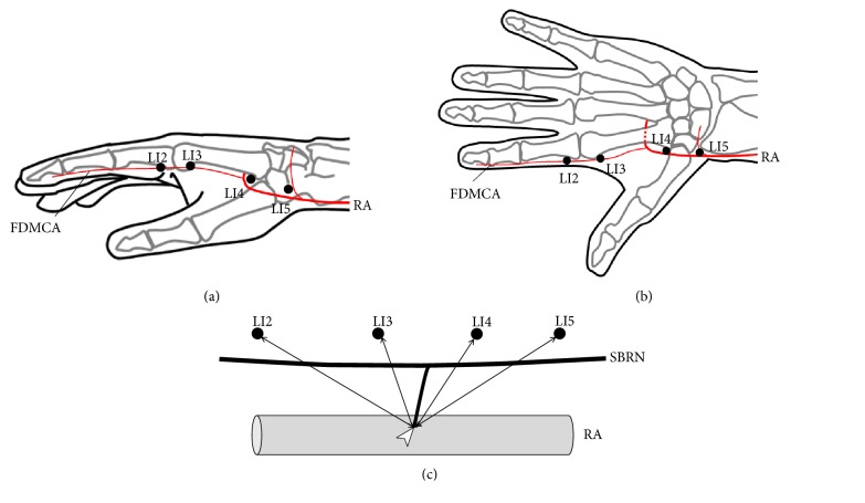
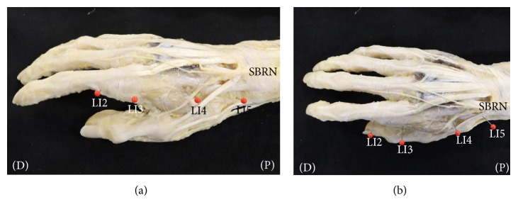
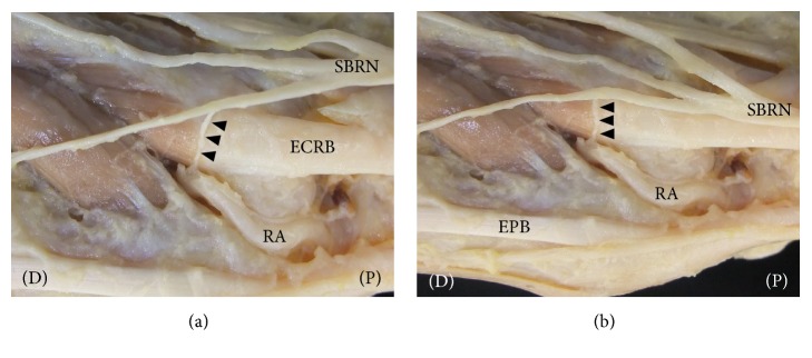
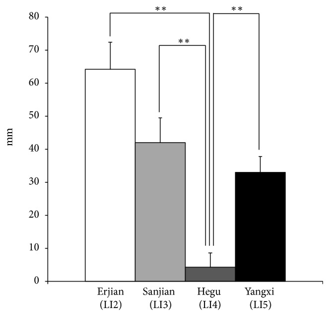
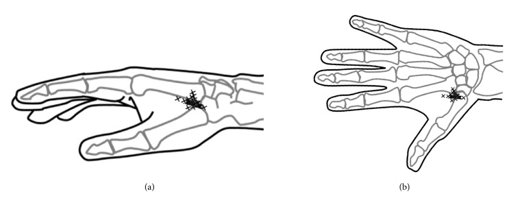
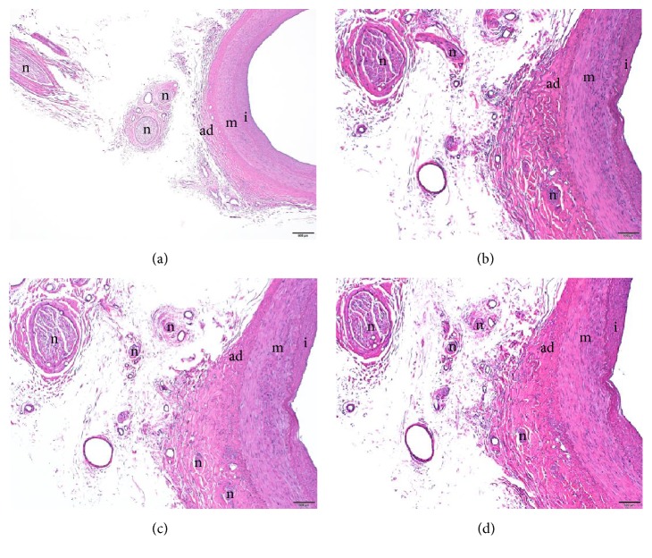
The acupuncture point "Hegu" (LI4) has been used for treating peripheral circulatory failure, which is located in the area covered by the superficial branch of the radial nerve (SBRN). SBRN has branches reaching arteries, so-called vascular branches (VBs), which are thought to be involved in the arterial constriction. The distribution areas of the VBs from the SBRN have been reported, but the positional relationship between these distribution areas and the acupuncture points are not known. To examine the positional relationship between LI4 and VBs from the SBRN, forty hands were examined to assess the positional relationship between the acupuncture points "Erjian" (LI2), "Sanjian" (LI3), LI4, and "Yangxi" (LI5) in the Yangming Large Intestine Meridian of Hand, which are located in the area covered by SBRN, and the VBs from the SBRN. After the VBs were identified, the distances from the acupuncture points (LI2, LI3, LI4, and LI5) to the point where the VBs reached the radial artery or the first dorsal metacarpal artery were measured. VBs reaching the radial arteries were observed in all specimens. The mean distances from LI2, LI3, LI4, and LI5 to the point where the VBs reached the radial artery were 64.2 ± 8.2 mm, 42.0 ± 7.5 mm, 4.3 ± 4.3 mm, and 33.0 ± 4.8 mm, respectively. LI4 was significantly closer than the other acupuncture points (P<0.01). The nerve fibers of the VBs adjacent to the radial artery were confirmed using hematoxylin and eosin staining. Our findings provide anatomical evidence that stimulation at LI4 is used for treating peripheral circulatory failure such as Raynaud's disease. LI4 is significant because it is located at a source point, making it clinically important.


Figures

Figure 1
Figure 1
Diagrams of anatomical measurements. (a) Schematic of acupuncture points in the Yangming Large Intestine Meridian of Hand on the radial side. (b) Schematic of acupuncture points in the Yangming Large Intestine Meridian of Hand on the dorsal side. (c) Method for measuring vascular branches. LI2, acupuncture point “Erjian”; LI3, acupuncture point “Sanjian”; LI4, acupuncture point “Hegu”; LI5, acupuncture point “Yangxi”; RA, radial artery; FDMCA, first dorsal metacarpal artery; SBRN, superficial branch of radial nerve. White arrowheads indicate the points where the VBs reached the arteries.
Figure 2
Figure 2
Dissection photographs showing the acupuncture points in the Yangming Large Intestine Meridian of Hand and the distribution of the superficial branch of the radial nerve (SBRN) on the right side. (a) Dissection photographs on the radial side. (b) Dissection photographs on the dorsal side; LI2, acupuncture point “Erjian”; LI3, acupuncture point “Sanjian”; LI4, acupuncture point “Hegu”; LI5, acupuncture point “Yangxi”; SBRN, superficial branch of radial nerve; (P), proximal (D), distal. The acupuncture points in the Yangming Large Intestine Meridian of Hand and the SBRN were identified after removal of the skin and subcutaneous tissue.
Figure 3
Figure 3
Dissection photographs showing the vascular branches (VBs) of the superficial branch of the radial nerve (SBRN) on the right side. (a) Dissection photographs on the radial side after removal of the skin and subcutaneous tissue. (b) Dissection photographs on the dorsal side after removal of the skin and subcutaneous tissue. RA, radial artery; SBRN, superficial branch of radial nerve; ECRB, extensor carpi radialis brevis tendon; EPB, extensor pollicis brevis muscle. Black arrowheads indicate the vascular branches (VBs). (P), proximal (D), distal.
Figure 4
Figure 4
The mean distances between the points where the VBs reached the radial artery and the positions of acupuncture points in the Yangming Large Intestine Meridian of Hand. LI4 was significantly closer to the point where the VB reached the radial artery than were LI2, LI3, and LI5 (Dunnett test, P<0.01). ∗∗P < 0.01, vs. LI4. Bars represent means with standard deviations (SD).
Figure 5
Figure 5
Diagrams showing the distribution of the points where the VBs reached the radial artery. (a) Schematic of the distribution of the points where the VBs reached the radial artery on the radial side. (b) Schematic of the distribution of the points where the VBs reached the radial artery on the dorsal side. Positions marked with an X indicate the points where the VBs reached the radial artery.
Figure 6
Figure 6
A vascular branch of the superficial branch of the radial nerve (SBRN). The nerve fibers of the vascular branch were adjacent to the radial artery (hematoxylin eosin stain). m, media; ad, adventitia; i, intima; n, nerve fibers of the vascular branch. All scale bars indicate 500 μm (Panel (a) ×2/0.16 objective lens; Panel (b)-(d) ×4/0.16 objective lens).

Comment in

References

    1. Sandberg M., Lundeberg T., Lindberg L., Gerdle B. Effects of acupuncture on skin and muscle blood flow in healthy subjects. European Journal of Applied Physiology. 2003;90(1-2):114–119. doi: 10.1007/s00421-003-0825-3. - DOI - PubMed
    1. Kramer J. G., Todd T. W. The distribution of nerves to the arteries of the arm, with a discussion of the clinical values of results. The Anatomical Record. 1914;8(5):243–255. doi: 10.1002/ar.1090080502. - DOI
    1. Sheehan D. On the innervation of the blood-vessels of the upper extremity: Some anatomical considerations. British Journal of Surgery. 1933;20(79):412–424. doi: 10.1002/bjs.1800207906. - DOI
    1. Pick J. The innervation of the arteries in the upper limb of man. The Anatomical Record. 1958;130(1):103–123. doi: 10.1002/ar.1091300109. - DOI - PubMed
    1. Pick J. The Autonomic Nervous System: Morphological, Comparative, Clinical, and Surgical Aspects. Philadelphia, PA, USA: J.B. Lippincott Co; 1970.

LinkOut - more resources