Case Reports
. 2019 Jun 23:2019:2719364.
doi: 10.1155/2019/2719364. eCollection 2019.

Diabetes Mellitus, Extreme Insulin Resistance, and Hypothalamic-Pituitary Langerhans Cells Histiocytosis

Affiliations
Case Reports

Diabetes Mellitus, Extreme Insulin Resistance, and Hypothalamic-Pituitary Langerhans Cells Histiocytosis

Mathilde Sollier et al. Case Rep Endocrinol. .

Abstract

Background: Langerhans Cell Histiocytosis (LCH) is a rare inflammatory neoplasm characterized by an infiltration of organs by Langerin + (CD207+) and CD1a+ histiocytes. Diabetes insipidus is a frequent manifestation of the disease, while diabetes mellitus is very rare. We report the first case of a 20-year-old man suffering from hypothalamopituitary histiocytosis and diabetes mellitus with serum anti-insulin receptor antibodies.

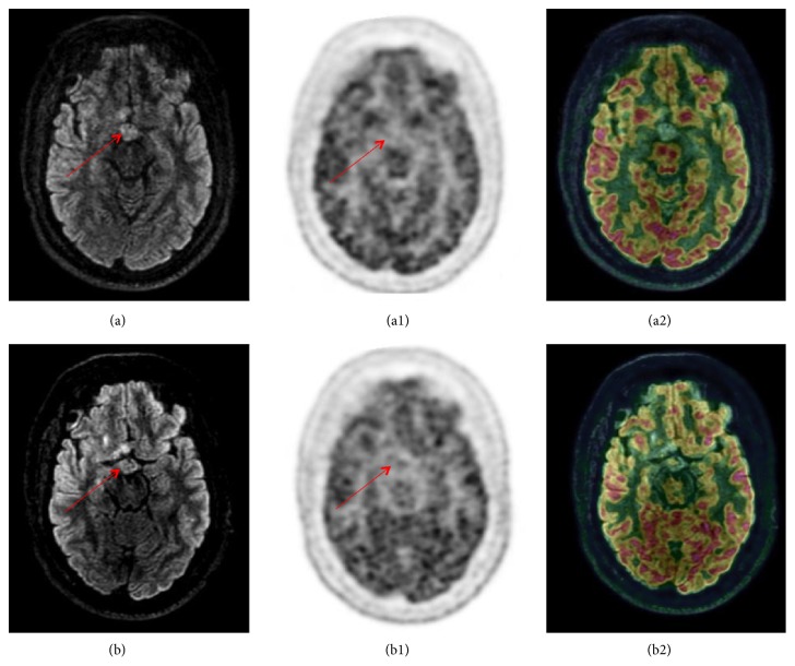
Case presentation: A 20-year-old patient was admitted for the evaluation of growth delay and hyperphagia. HbA1c level and fasting blood glucose were in the normal range. The diagnosis of hypothalamopituitary histiocytosis was based on histological features after biopsy of a large suprachiasmatic lesion identified on magnetic resonance imaging (MRI). Association of vinblastine and purinethol was started followed by a second-line therapy by cladribine. During the follow-up, the patient was admitted for recurrence of hyperglycemic states and extreme insulin resistance. The screening for serum anti-insulin receptor antibodies was positive. Each episode of hyperglycemia appeared to be correlated with tumoral activity and increase in serum anti-insulin receptor antibodies and appeared to be improved when the disease was controlled by chemotherapy.

Conclusion: We report the first description of a hypothalamopituitary histiocytosis associated with serum anti-insulin receptor antibodies, extreme insulin resistance, and diabetes. Parallel evolution of glucose levels and serum anti-insulin receptor antibodies seemed to be the consequence of immune suppressive properties of cladribine.


Figures

Figure 1
Figure 1
Evolution of brain MRI during follow-up. At diagnosis, the normal T1-weighted hyperintense signal of the posterior pituitary has disappeared (a). Contrast-enhanced hypothalamic mass after gadolinium infusion ((b) and (c)); compression of third ventricle and thickening of pituitary stalk (b). Comparison of brain MRI (T1 with gadolinium) at diagnosis (M0) (d) and at the end of the first-line therapy (e). MRI showed a significant decrease of the hypothalamic mass but the persistence of contrast enhancement after gadolinium. Brain MRI (T1 after gadolinium infusion) at M30 showing a hypothalamic lesion stable in size and with decreased hypersignal compared to previous MRI (f).
Figure 2
Figure 2
Sagittal PET-MRI brain imaging. Sagittal PET-MRI brain imaging with [18F]FDG at M32 (a, a1, a2) and at M35 (b, b1, b2) of follow-up. Nodular thickening of the floor of the third ventricle and pituitary infundibulum, in hyperintense FLAIR, without [18F]FDG-uptake, measuring 7 mm in height x 13 mm in the axial plane, without significant enhancement, globally unchanged from the previous MRI. Linear and fine enhancement of the pituitary infundibulum with stable appearance.
Figure 3
Figure 3
Axial PET-MRI brain imaging. Axial PET-MRI brain imaging with [18F]FDG at M32 (a, a1, a2) and at M35 (b, b1, b2) of follow-up.

References

    1. Emile J.-F., Abla O., Fraitag S., et al. Revised classification of histiocytoses and neoplasms of the macrophage-dendritic cell lineages. Blood. 2016;127(22):2672–2681. doi: 10.1182/blood-2016-01-690636. - DOI - PMC - PubMed
    1. Haroche J., Cohen-Aubart F., Rollins B. J., et al. Histiocytoses: emerging neoplasia behind inflammation. The Lancet Oncology. 2017;18(2):e113–e125. doi: 10.1016/S1470-2045(17)30031-1. - DOI - PubMed
    1. Guyot-Goubin A., Donadieu J., Barkaoui M., Bellec S., Thomas C., Clavel J. Descriptive epidemiology of childhood Langerhans cell histiocytosis in France, 2000-2004. Pediatric Blood & Cancer. 2008;51(1):71–75. doi: 10.1002/pbc.21498. - DOI - PubMed
    1. The Writing Group of the Histiocyte Society. Histiocytosis syndromes in children. The Lancet. 1987;329(8526):208–209. doi: 10.1016/s0140-6736(87)90016-x. - DOI - PubMed
    1. Makras P., Alexandraki K. I., Chrousos G. P., Grossman A. B., Kaltsas G. A. Endocrine manifestations in Langerhans cell histiocytosis. Trends in Endocrinology & Metabolism. 2007;18(6):252–257. doi: 10.1016/j.tem.2007.06.003. - DOI - PubMed

Publication types

LinkOut - more resources