Review
. 2019 Jun;9(3):250-255.
doi: 10.1212/CPJ.0000000000000605.

Acute middle cerebral artery stroke in a patient with a patent middle cerebral artery

Affiliations
Review

Acute middle cerebral artery stroke in a patient with a patent middle cerebral artery

Jamie Cooke et al. Neurol Clin Pract. 2019 Jun.

Abstract

Purpose of review: Knowledge of cerebrovascular anatomical variants is vital for clinicians working with patients presenting with signs and symptoms of cerebral infarction, particularly in the era of endovascular clot retrieval.

Recent findings: We provide an overview of a cerebrovascular anatomical variation and detail a patient presenting with cerebral infarction secondary to occlusion of their anomalous vessel who underwent successful endovascular clot retrieval with excellent functional outcome. We also include technical descriptions.

Summary: Given the clinical importance of the areas supplied by the accessory middle cerebral artery, knowledge of this vessel is not only important for diagnosis but also for neurosurgical or endovascular management of patients with this variant.


Figures

Figure 1
Figure 1. Imaging on presentation
(A) Noncontrast brain CT showing established ischemic changes within the frontal lobe in the left middle cerebral artery territory (red arrow). (B) Noncontrast brain CT with a hyperattenuating intravascular thrombus (green arrow) on the left correlating with the expected position of a left middle cerebral artery thrombus. (C) Tmax and (D) cerebral blood volume from a CT perfusion demonstrating extensive ischemic penumbra of the left MCA territory with suggestion of ischemic mismatch. (E and F) Maximum intensity projection from CT angiography demonstrates normal contrast opacification in a horizontal middle cerebral arterial vessel and the distal cortical branches of the middle cerebral artery.
Figure 2
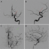
Figure 2. Digital subtraction angiography via left internal carotid artery injection
Digital subtraction angiography via left internal carotid artery injection. (A–D) Irregularity (red arrows) of the proximal A1 segment of the anterior cerebral artery was noted, as well as a lack of filling within the superior division cortical branches, and lenticulostriate vessels in the middle cerebral artery territory (green arrow) correlating with findings on CT angiography. This also confirmed patency of a horizontal middle cerebral arterial vessel and the distal cortical branches as identified on the CT angiography.
Figure 3
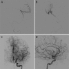
Figure 3. Digital subtraction angiography during/after procedure
(A and B) After gentle advancement of a Synchro2 microwire, the abnormality was felt to relate to an accessory middle cerebral artery. A Trevo microcatheter was advanced beyond the occlusion into distal branches of the accessory middle cerebral artery correlating with suspected missing branches on the initial angiogram. (C and D) After a single pass with a 4 × 40-mm Solitaire Stentriever, all middle cerebral artery branches opacified with mTICI 3 recanalization. Note increased opacification of perforating lenticulostriate arteries to the deep white and gray matter structures.
Figure 4
Figure 4. Follow-up MRI 3 months after ECR
(A) T2-weighted image showing minor left-sided basal ganglia infarction. (B) FLAIR image showing no cortical infarction. FLAIR = fluid attenuation inversion recovery.

Comment in

References

    1. Crompton MR. The pathology of ruptured middle-cerebral aneurysms with special reference to the differences between the sexes. Lancet 1962;2:421–425. - PubMed
    1. Komiyama M, Nakajima H, Nishikawa M, Yasui T. Middle cerebral artery variations: duplicated and accessory arteries. AJNR Am J Neuroradiol 1998;19:45–49. - PMC - PubMed
    1. Abanou A, Lasjaunias P, Manelfe C, Lopez-Ibor L. The accessory middle cerebral artery (AMCA): diagnostic and therapeutic consequences. Anat Clin 1984;6:305–309. - PubMed
    1. Kim MS, Lee HK. The angiographic feature and clinical implication of accessory middle cerebral artery. J Korean Neurosurg Soc 2009;45:289–292. - PMC - PubMed
    1. Marinkovic S, Milisavljevic M, Kovacevic M. Anatomical bases for surgical approach to the initial segment of the anterior cerebral artery: microanatomy of Heubner's artery and perforating branches of the anterior cerebral artery. Surg Radiol Anat 1986;8:7–18. - PubMed