

The clinical features of posterior scleritis with serous retinal detachment: a retrospective clinical analysis
- PMID: 31341807
- PMCID: PMC6629818
- DOI: 10.18240/ijo.2019.07.16
The clinical features of posterior scleritis with serous retinal detachment: a retrospective clinical analysis
Abstract
Aim: To summarize the clinical features, systemic associations, risk factors and choroidal thickness (CT) changing in posterior scleritis (PS) with serous retinal detachment.
Methods: This retrospective study included 23 patients diagnosed PS with retinal detachment from August 2012 to July 2017. All patients' medical history and clinical features were recorded. The examinations included best corrected visual acuity (BCVA), intraocular pressure (IOP), fundus examination, and routine eye examinations. Posterior coats thickness (PCT) was determined by B-scan ultrasound, the CT was measured by enhanced depth imaging spectral-domain optical coherence tomography (EDI-OCT) and clinical data were compiled and analyzed.
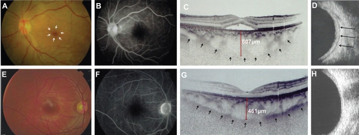
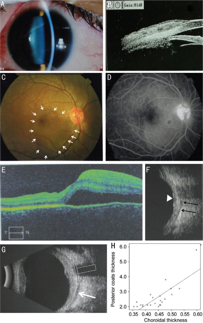
Results: After application of extensive exclusion criteria, 23 patients with PS remained (13 females, 10 males). The average age at presentation was 29.5±9.24 years old. Ocular pain and blurred vision were the two most common complained symptoms by patients. Anterior scleritis occurred in 12 patients, which was confirmed by ultrasound biomicroscopy (UBM) examination. Despite all patients displaying serous retinal detachment in their macula, no fluorescein leakage was observed in the macular area. Optic disc swelling was documented in 10 of the 23 eyes. From B-scan ultrasound examination, the PCT increased with fluid in Tenon's capsule demonstrated as a typical T-sign. The average PCT was 2.51±0.81 mm in the PS-affected eyes and only 1.09±0.29 mm in the unaffected eye (P<0.0001). The subfoveal CT was 442.61±55.61 µm, which correlated with axis length (r=-0.65, P=0.001) and PCT (r=0.783, P<0.001). The BCVA and IOP did not correlate with either CT or PCT.
Conclusion: PS with serous retinal detachment presented a variety of symptoms, such as pain, visual loss, and physical indicators. Typical T-sign detected by B-scan ultrasound is a useful confirmatory sign for PS diagnosis. Pathological increases in CT might be a potential predictive factor for inflammation.
Keywords: choroidal thickness; clinical features; posterior scleritis; scleritis; serous retinal detachment.
Figures
References
-
- Biswas J, Mittal S, Ganesh SK, Shetty NS, Gopal L. Posterior scleritis: clinical profile and imaging characteristics. Indian J Ophthalmol. 1998;46(4):195–202. - PubMed
-
- Yang P, Ye Z, Tang J, Du L, Zhou Q, Qi J, Liang L, Wu L, Wang C, Xu M, Tian Y, Kijlstra A. Clinical features and complications of scleritis in Chinese patients. Ocul Immunol Inflamm. 2018;26(3):387–396. - PubMed
-
- Lavric A, Gonzalez-Lopez JJ, Majumder PD, Bansal N, Biswas J, Pavesio C, Agrawal R. Posterior scleritis: analysis of epidemiology, clinical factors, and risk of recurrence in a cohort of 114 patients. Ocul Immunol Inflamm. 2016;24(1):6–15. - PubMed
LinkOut - more resources
Full Text Sources