Review
. 2019 Dec;46(13):2848-2858.
doi: 10.1007/s00259-019-04426-0. Epub 2019 Jul 24.

Engineered antibodies: new possibilities for brain PET?

Collaborators, Affiliations
Review

Engineered antibodies: new possibilities for brain PET?

Dag Sehlin et al. Eur J Nucl Med Mol Imaging. 2019 Dec.

Abstract

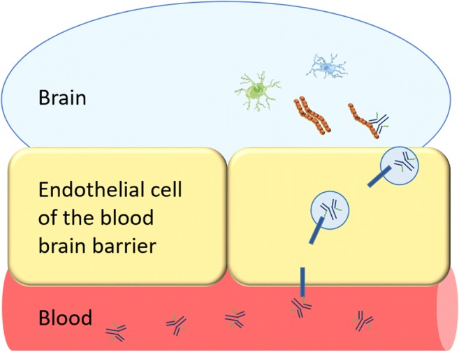
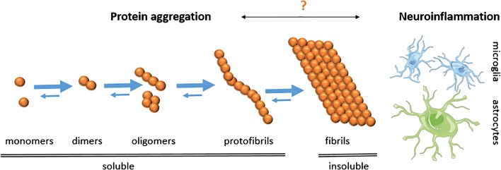
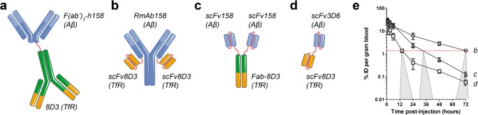
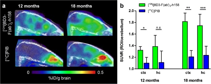
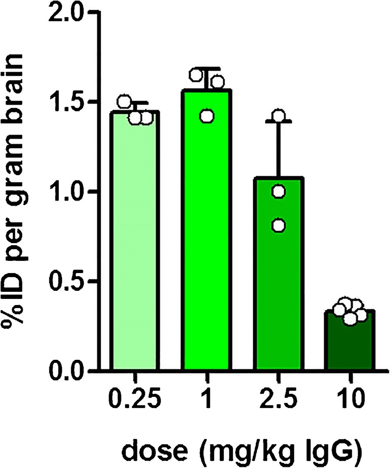
Almost 50 million people worldwide are affected by Alzheimer's disease (AD), the most common neurodegenerative disorder. Development of disease-modifying therapies would benefit from reliable, non-invasive positron emission tomography (PET) biomarkers for early diagnosis, monitoring of disease progression, and assessment of therapeutic effects. Traditionally, PET ligands have been based on small molecules that, with the right properties, can penetrate the blood-brain barrier (BBB) and visualize targets in the brain. Recently a new class of PET ligands based on antibodies have emerged, mainly in applications related to cancer. While antibodies have advantages such as high specificity and affinity, their passage across the BBB is limited. Thus, to be used as brain PET ligands, antibodies need to be modified for active transport into the brain. Here, we review the development of radioligands based on antibodies for visualization of intrabrain targets. We focus on antibodies modified into a bispecific format, with the capacity to undergo transferrin receptor 1 (TfR1)-mediated transcytosis to enter the brain and access pathological proteins, e.g. amyloid-beta. A number of such antibody ligands have been developed, displaying differences in brain uptake, pharmacokinetics, and ability to bind and visualize the target in the brain of transgenic mice. Potential pathological changes related to neurodegeneration, e.g. misfolded proteins and neuroinflammation, are suggested as future targets for this novel type of radioligand. Challenges are also discussed, such as the temporal match of radionuclide half-life with the ligand's pharmacokinetic profile and translation to human use. In conclusion, brain PET imaging using bispecific antibodies, modified for receptor-mediated transcytosis across the BBB, is a promising method for specifically visualizing molecules in the brain that are difficult to target with traditional small molecule ligands.

Keywords: Alzheimer’s disease (AD); Amyloid-β (Aβ); Antibody; Blood–brain barrier (BBB); Positron emission tomography (PET); Transferrin receptor 1 (TfR1)-mediated transcytosis.


Conflict of interest statement

The authors declare they have no conflict of interest.

Figures

Fig. 1
Fig. 1
Transferrin receptor (TfR)-mediated transcytosis of a bispecific antibody. The bispecific antibody binds to TfR at the luminal side of the BBB. TfR transports the bispecific antibody inside an endosome across the BBB. The bispecific antibody is released on the abluminal side of the BBB and can then bind to its target (e.g. Aβ, reactive astrocytes, microglia) in the brain parenchyma
Fig. 2
Fig. 2
Protein misfolding causes aggregation of monomeric Aβ into oligomers and protofibrils, which ultimately form insoluble fibrils, i.e. the main constituent of Aβ plaques which are characteristic for AD. Of the soluble species, oligomers/protofibrils are most likely the toxic forms of Aβ responsible for neurodegeneration. However, all amyloid PET ligands today detect fibrillar Aβ. The trigger for neuroinflammation is debated, but both activated microglia and reactive astrocytes are observed in the AD brain
Fig. 3
Fig. 3
Aβ pathology visualized in the transgenic (tg-ArcSwe) mouse brain. Ex vivo autoradiography brain sections isolated 6 days after intravenous administration of (a) a bispecific TfR1- and Aβ-binding antibody or (b) an unmodified Aβ-binding antibody. The spatial distribution of the bispecific antibody corresponds well with the Aβ40 immunohistochemistry shown in (c), while the non-modified antibody is accumulated centrally around the ventricles and in high-intensity deposits
Fig. 4
Fig. 4
Sagittal PET images obtained at 3 days after administration of the bispecific radioligand [124I]8D3-F(ab’)2-h158 in two mouse models of AD (ArcSwe and Swe) and wild-type (WT) mice at different ages
Fig. 5
Fig. 5
Different formats of bispecific, brain-penetrating antibodies used for PET imaging of Aβ pathology in AD transgenic mouse models. a F(ab’)2 fragment of humanized Aβ protofibril-selective mAb158, chemically coupled to full 8D3 antibody (Mw ~270 kDa). b Recombinant variant of mAb158 (RmAb158) with scFv8D3 recombinantly fused to the C terminus of each of the light chains (Mw ~210 kDa). c Tribody composed of two scFv158 attached to each chain of a Fab-8D3, brought together by the natural combination of the Fab fragment (Mw ~110 kDa). d Tandem-scFv composed of scFv3D6 fused via a polypeptide linker to scFv8D3 (Mw ~58 kDa). e Blood elimination curves of recombinant antibody ligands (b), (c), and (d). The dashed red line represents an approximate antibody blood concentration threshold below which PET imaging is feasible. From the intersection of each antibody’s blood curve with the threshold line, there is a projection to a time window where AD transgenic mice can at the earliest be discriminated from wild-type mice
Fig. 6
Fig. 6
Comparison of antibody and PIB PET imaging. a Sagittal view of PET images obtained from 12- and 18-month-old tg-ArcSwe mice 3 days post-injection of [124I]8D3-F(ab’)2-h158 (upper panel) in comparison with mice scanned 40–60 min after injection of [11C]PIB (lower panel). b Quantification of PET images from (a) expressed as standardized uptake value ratio (SUVR) of two regions of interest, cortex (ctx) and hippocampus (hc), using cerebellum as reference region. Significant differences were observed for all regions and ages except for hippocampus in the 12-month-old animals
Fig. 7
Fig. 7
Dose vs brain uptake of RmAb158-scFv8D3. Brain antibody uptake (% of injected dose per gram brain tissue) was measured 2 h post-injection of RmAb158-scFv8D3 at doses ranging from 0.25 mg/kg body weight to a high therapeutic dose of 10 mg/kg body weight. While antibody doses relevant to PET imaging (below 0.5 mg/kg) had no impact on brain delivery, doses above 1 mg/kg seemed to saturate the TfR transport mechanism, resulting in reduced brain uptake

References

    1. Niemeijer AN, et al. Whole body PD-1 and PD-L1 positron emission tomography in patients with non-small-cell lung cancer. Nat Commun. 2018;9(1):4664. - PMC - PubMed
    1. Jauw YW, et al. Immuno-positron emission tomography with Zirconium-89-labeled monoclonal antibodies in oncology: what can we learn from initial clinical trials? Front Pharmacol. 2016;7:131. - PMC - PubMed
    1. Bard F, et al. Peripherally administered antibodies against amyloid beta-peptide enter the central nervous system and reduce pathology in a mouse model of Alzheimer disease. Nat Med. 2000;6(8):916–919. - PubMed
    1. Poduslo JF, Curran GL, Berg CT. Macromolecular permeability across the blood-nerve and blood-brain barriers. Proc Natl Acad Sci U S A. 1994;91(12):5705–5709. - PMC - PubMed
    1. Pardridge WM. Re-engineering therapeutic antibodies for Alzheimer's disease as blood-brain barrier penetrating bi-specific antibodies. Expert Opin Biol Ther. 2016;16(12):1455–1468. - PubMed