

Parallel Architecture of Fully Convolved Neural Network for Retinal Vessel Segmentation
- PMID: 31342298
- PMCID: PMC7064708
- DOI: 10.1007/s10278-019-00250-y
Parallel Architecture of Fully Convolved Neural Network for Retinal Vessel Segmentation
Abstract
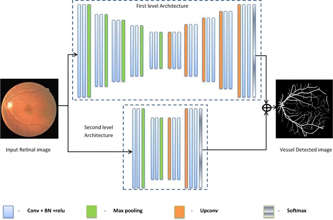
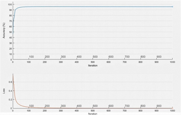
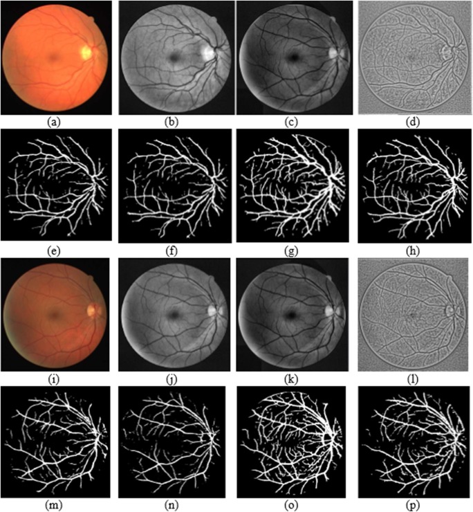
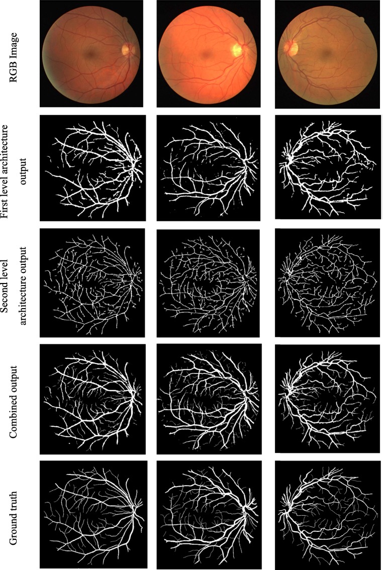
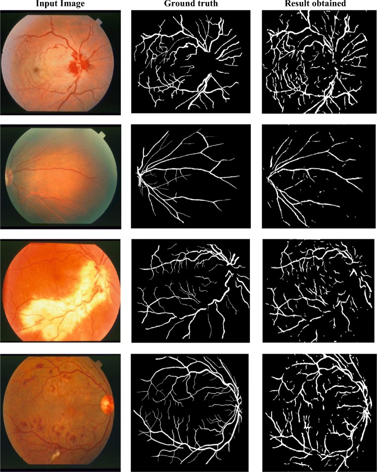
Retinal blood vessel extraction is considered to be the indispensable action for the diagnostic purpose of many retinal diseases. In this work, a parallel fully convolved neural network-based architecture is proposed for the retinal blood vessel segmentation. Also, the network performance improvement is studied by applying different levels of preprocessed images. The proposed method is experimented on DRIVE (Digital Retinal Images for Vessel Extraction) and STARE (STructured Analysis of the Retina) which are the widely accepted public database for this research area. The proposed work attains high accuracy, sensitivity, and specificity of about 96.37%, 86.53%, and 98.18% respectively. Data independence is also proved by testing abnormal STARE images with DRIVE trained model. The proposed architecture shows better result in the vessel extraction irrespective of vessel thickness. The obtained results show that the proposed work outperforms most of the existing segmentation methodologies, and it can be implemented as the real time application tool since the entire work is carried out on CPU. The proposed work is executed with low-cost computation; at the same time, it takes less than 2 s per image for vessel extraction.
Keywords: Deep learning; Enhancement; Fully convolved neural network; Retinal vessel.
Conflict of interest statement
The authors declare that they have no conflict of interest.
Figures


References
-
- Lupas¸cu CA, Tegolo D, Trucco E. FABC: retinal vessel segmentation using AdaBoost. IEEE Trans Inf Technol Biomed. 2010;14(5):1267–1274. - PubMed
-
- Dizdaroglu B, Cansizoglu E a, Kalpathy-Cramer J, keck K, Chiang MF, Erdogmus D. Structure based level set method for automatic retinal vasculature segmentation. EURASIP J Image Video Process. 2014;1(39):1–26.
-
- Barkhoda W, Akhlaqian F, Amiri MD, Nouroozzadeh MS. Retina identification based on the pattern of blood vessels using fuzzy logic. EURASIP J Adv Signal Process. 2011;1(113):1–8.
-
- Zhao J, Yang J, Ai D, HongSong Y, Jiang YH, Luosh Z, Wang Y. Automatic retinal vessel segmentation using multi-scale superpixel chain tracking. Digital Signal Process. 2018;81:26–41.
-
- Marin D, Aquino A, Arias MEG, Bravo JM. A new supervised method for blood vessel segmentation in retinal images by using gray-level and moment invariants-based features. IEEE Trans Med Imaging. 2011;30(1):146–158. - PubMed
MeSH terms
LinkOut - more resources
Full Text Sources
Medical
