. 2019 Oct;110(10):3340-3349.
doi: 10.1111/cas.14142. Epub 2019 Aug 13.

Impaired ligand-dependent MET activation caused by an extracellular SEMA domain missense mutation in lung cancer

Affiliations

Impaired ligand-dependent MET activation caused by an extracellular SEMA domain missense mutation in lung cancer

Wenyu Miao et al. Cancer Sci. 2019 Oct.

Abstract

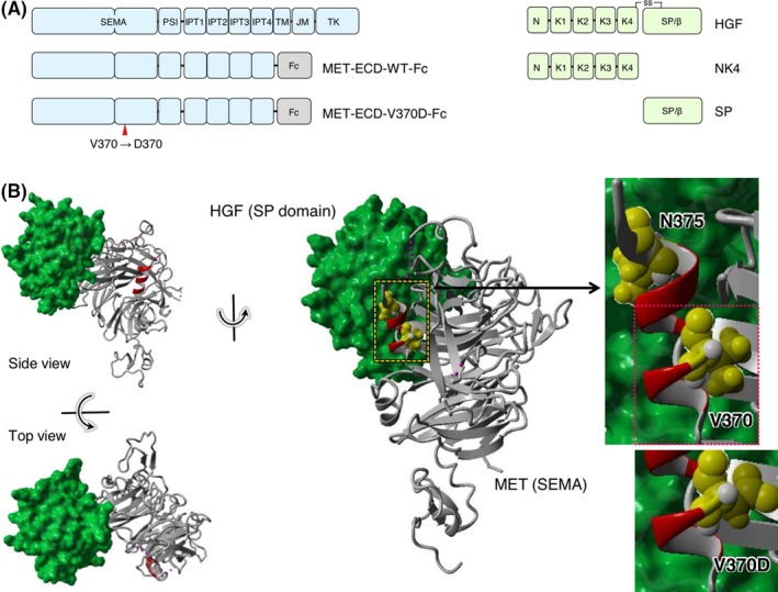
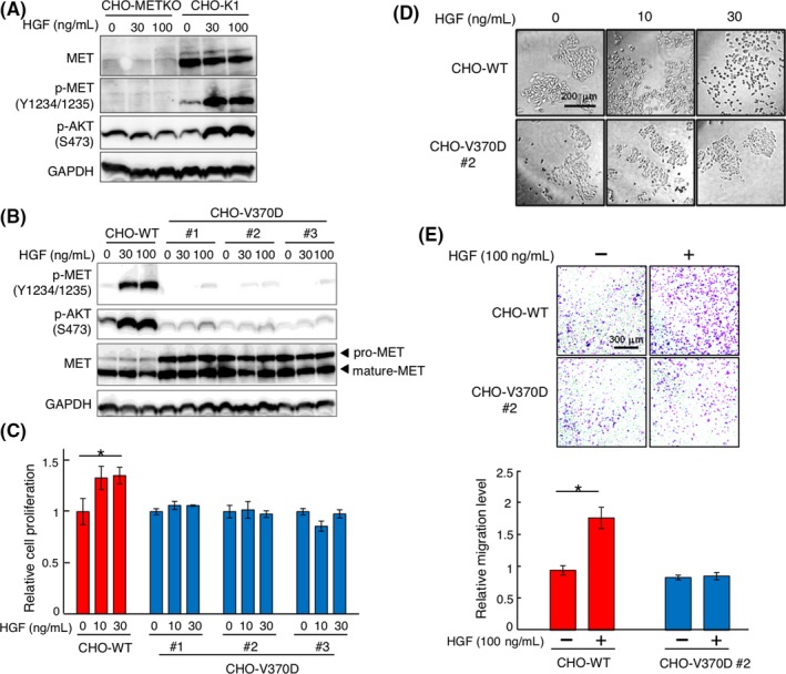
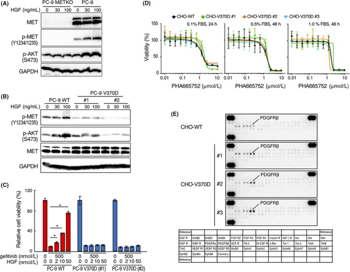
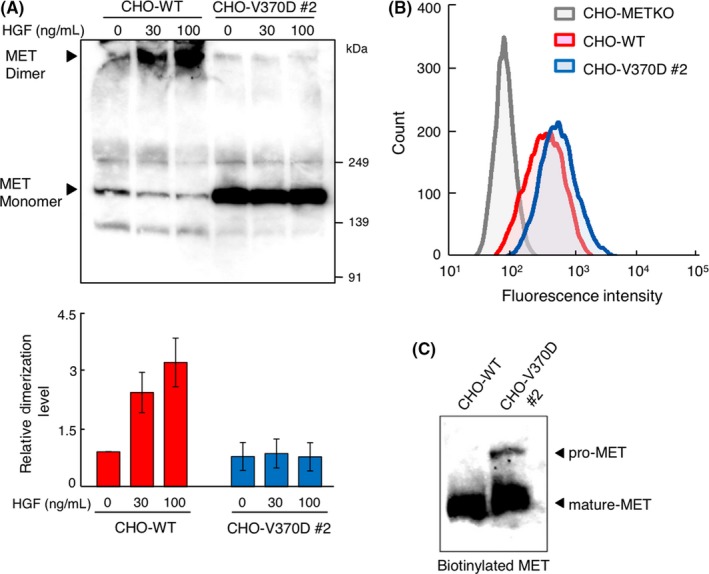
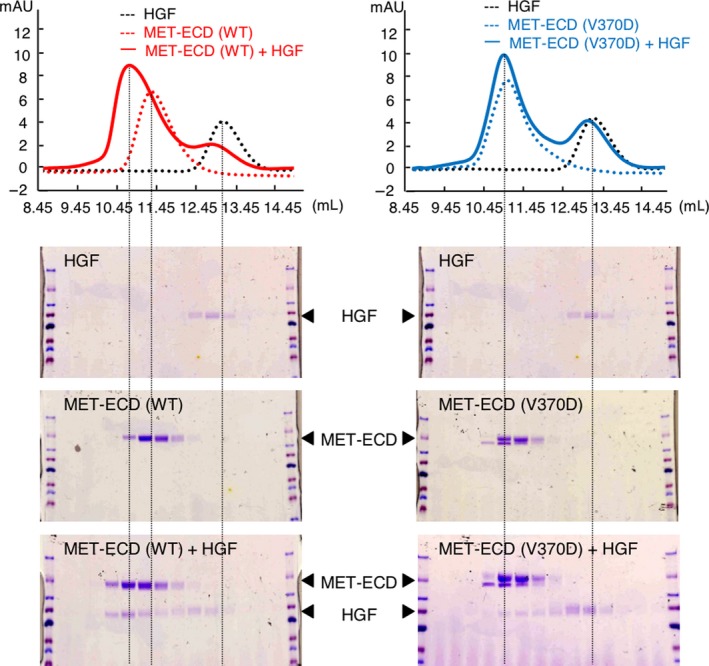
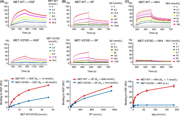
Aberrant activation of the MET/hepatocyte growth factor (HGF) receptor participates in the malignant behavior of cancer cells, such as invasion-metastasis and resistance to molecular targeted drugs. Many mutations in the MET extracellular region have been reported, but their significance is largely unknown. Here, we report the dysregulation of mutant MET originally found in a lung cancer patient with Val370 to Asp370 (V370D) replacement located in the extracellular SEMA domain. MET-knockout cells were prepared and reconstituted with WT-MET or V370D-MET. HGF stimulation induced MET dimerization and biological responses in cells reconstituted with WT-MET, but HGF did not induce MET dimerization and failed to induce biological responses in V370D-MET cells. The V370D mutation abrogated HGF-dependent drug resistance of lung cancer cells to epidermal growth factor receptor-tyrosine kinase inhibitors (EGFR-TKI). Compared with WT-MET cells, V370D-MET cells showed different activation patterns in receptor tyrosine kinases upon exposure to survival/growth-stressed conditions. Surface plasmon resonance analysis indicated that affinity between the extracellular region of V370D-MET and HGF was reduced compared with that for WT-MET. Further analysis of the association between V370D-MET and the separate domains of HGF indicated that the SP domain of HGF was unchanged, but its association with the NK4 domain of HGF was mostly lost in V370D-MET. These results indicate that the V370D mutation in the MET receptor impairs the functional association with HGF and is therefore a loss-of-function mutation. This mutation may change the dependence of cancer cell growth/survival on signaling molecules, which may promote cancer cell characteristics under certain conditions.

Keywords: MET receptor; affinity; hepatocyte growth factor; loss of function; missense mutation.


Conflict of interest statement

Authors declare no conflicts of interest for this article.

Figures

Figure 1
Figure 1
Structures of hepatocyte growth factor (HGF) and the MET receptor. A, Domain architecture of MET, HGF, and their recombinant domains. B, Complex structure composed of the SEMA domain of MET and the SP domain of HGF (PDB number: 1SHY)
Figure 2
Figure 2
Hepatocyte growth factor (HGF)‐induced responses in CHOWT and CHO‐V370D cells. A, Activation of MET and AKT in CHO‐K1 and CHO‐K1 MET knockout (CHOMETKO) cells. Cells were stimulated with HGF for 10 min. MET, p‐MET, p‐AKT, and GAPDH were detected by western blotting. B, Expression and activation of METWT and MET‐V370D in CHOMETKO cells and response to HGF. Cells were stimulated with HGF for 10 min. MET, p‐MET, p‐AKT, and GAPDH were detected by western blotting. C, Cell proliferation induced by HGF. Cells were cultured with or without HGF for 48 h. Each value indicates the mean ± SE of triplicate measurements. Asterisk indicates < .05. D, HGF‐induced cell scattering. Cells were cultured with or without HGF for 48 hours, representative images of which are shown. E, HGF‐induced cell migration. Cells were seeded on Transwell membranes and cultured for 24 h. Each value indicates the mean ± SE of triplicate measurements. Asterisk indicates < .05. Representative images are shown
Figure 3
Figure 3
Distinct responses mediated by WT and V370D mutant MET. A, Expression and tyrosine phosphorylation of MET receptor in PC‐9 MET receptor‐knockout (PC‐9 METKO) cells. Cells were stimulated with HGF for 10 min. MET, p‐MET, p‐AKT, and GAPDH were detected by western blotting. B, Expression and activation of METWT and MET‐V370D and response to HGF stimulation. Cells were stimulated with HGF for 10 min. MET, p‐MET, p‐AKT, and GAPDH were detected by western blotting. C, Cell viability after treatment with gefitinib and hepatocyte growth factor (HGF) in PC‐9 WT and PC‐9 V370D cells. Cells were treated with or without gefitinib or HGF for 3 d. Data represent mean ± SE (n = 3). *< .05. D, Effect of MET kinase inhibitor on viability of CHOWT and CHO‐V370D cells. Cells were cultured in the absence or presence of PHA665752 in medium supplemented with different FBS concentrations for 24 or 48 h. Data represent mean ± SD (n = 4). E, Change in phosphorylation of RTK in CHOWT cells and CHO‐V370D cells (#1‐#3, independent clones) cultured under survival/growth‐stressed conditions. The cells were cultured in medium supplemented with 0.01% FBS and 20 ng/mL HGF for 10 d
Figure 4
Figure 4
MET receptor dimerization and cell surface localization of V370D‐MET. A, MET receptor dimerization induced by hepatocyte growth factor (HGF). CHOWT and CHO‐V370D cells were stimulated with HGF followed by treatment with a cross‐linker bis(sulfosuccinimidyl)suberate (BS3). MET dimerization was analyzed by immunoprecipitation and western blotting using an anti‐MET antibody. Relative MET dimerization level was calculated by the band intensity in western blots. Each value indicates the mean ± SE of triplicate measurements. Representative image is shown. B, Cell surface expression of MET receptor. Cells were stained with FITC‐conjugated anti‐MET antibody and cell surface expression of MET was analyzed by flow cytometry. C, Cell surface MET receptor detected by biotinylation assay. Biotinylated cell surface proteins were pulled down, and cell surface MET was detected by western blotting using an anti‐MET antibody
Figure 5
Figure 5
Binding of hepatocyte growth factor (HGF) to METWT and MET‐V370D as evaluated by size‐exclusion chromatography. HGF and METECD‐His (WT or V370D) were subjected to size‐exclusion chromatography alone or as a mixture. Eluted fractions were analyzed by SDSPAGE and protein staining with Coomassie Brilliant Blue
Figure 6
Figure 6
Binding of hepatocyte growth factor (HGF), SP, and NK4 to METWT and MET‐V370D. Binding kinetics of HGF (A), SP (B), and NK4 (C) to METWT or MET‐V370D was measured by surface plasmon resonance (SPR) analysis. In (A), biotinylated HGF was immobilized on a sensor chip and binding of METECD‐Fc (WT or V370D) was measured (n = 2). In (B) and (C), METECD‐Fc‐His (WT or V370D) was immobilized on a sensor chip and binding of SP or NK4 was measured (n = 2)

References

    1. Park M, Dean M, Kaul K, Braun MJ, Gonda MA, Vande Woude G. Sequence of MET protooncogene cDNA has features characteristic of the tyrosine kinase family of growth‐factor receptors. Proc Natl Acad Sci USA. 1987;84:6379‐6383. - PMC - PubMed
    1. Comoglio PM, Trusolino L, Boccaccio C. Known and novel roles of the MET oncogene in cancer: a coherent approach to targeted therapy. Nat Rev Cancer. 2018;18:341‐358. - PubMed
    1. Komada M, Hatsuzawa K, Shibamoto S, Ito F, Nakayama K, Kitamura N. Proteolytic processing of the hepatocyte growth factor/scatter factor receptor by furin. FEBS Lett. 1993;328:25‐29. - PubMed
    1. Tempest PR, Stratton MR, Cooper CS. Structure of the met protein and variation of met protein kinase activity among human tumour cell lines. Br J Cancer. 1988;58:3‐7. - PMC - PubMed
    1. Gherardi E, Youles ME, Miguel RN, et al. Functional map and domain structure of MET, the product of the c‐met protooncogene and receptor for hepatocyte growth factor/scatter factor. Proc Natl Acad Sci. 2003;100:12039‐12044. - PMC - PubMed