Review
. 2019 Dec;198(3):314-325.
doi: 10.1111/cei.13353. Epub 2019 Aug 30.

Immune cell trafficking to the islets during type 1 diabetes

Affiliations
Review

Immune cell trafficking to the islets during type 1 diabetes

A M Sandor et al. Clin Exp Immunol. 2019 Dec.

Abstract

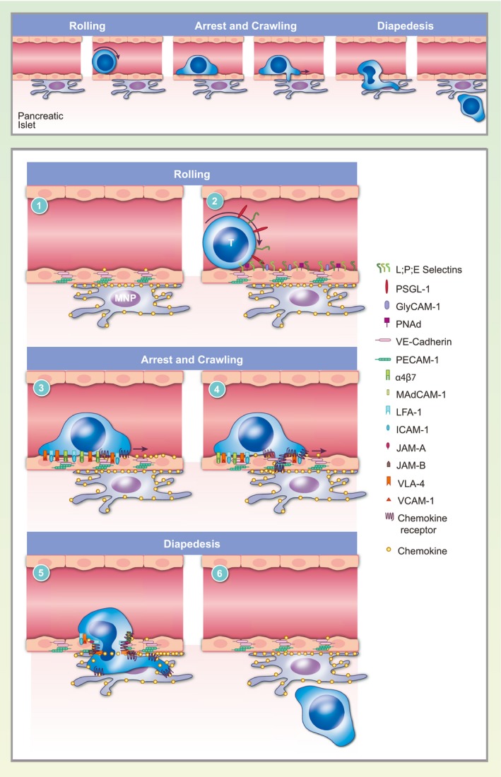
Inhibition of immune cell trafficking to the pancreatic islets during type 1 diabetes (T1D) has therapeutic potential, since targeting of T cell and B cell trafficking has been clinically effective in other autoimmune diseases. Trafficking to the islets is characterized by redundancy in adhesion molecule and chemokine usage, which has not enabled effective targeting to date. Additionally, cognate antigen is not consistently required for T cell entry into the islets throughout the progression of disease. However, myeloid cells are required to enable T cell and B cell entry into the islets, and may serve as a convergence point in the pathways controlling this process. In this review we describe current knowledge of the factors that mediate immune cell trafficking to pancreatic islets during T1D progression.

Keywords: adhesion molecules; autoimmunity; cell trafficking; chemokines; diabetes.


Figures

Figure 1
Figure 1
T cell extravasation into the pancreatic islets. The process of extravasation can be broken down into rolling on the vascular wall, firm adhesion and crawling and diapedesis. T cell extravasation is mediated by molecular interactions between the T cells and the vascular endothelium. (1) Interactions between vascular adhesion molecules, including vascular endothelial (VE)‐cadherin, platelet and endothelial cell adhesion molecule 1 (PECAM‐1) and junctional adhesion molecule (JAM) proteins, maintain tight junctions between endothelial cells forming the vascular barrier. Chemokine‐producing mononuclear phagocytes associate with the islet vasculature. (2) T cells roll on the vascular endothelium by tethering to P‐selectin glycoprotein ligand 1 (PSGL‐1) and L‐selectin through low affinity interactions between T cell‐expressed selectins and cell adhesion molecules on the vasculature. (3) Chemokine signaling causes T cells to arrest on the vascular endothelium by promoting the high‐affinity conformation of integrins such as lymphocyte function‐associated antigen 1 (LFA‐1), very late antigen 4 (VLA‐4) and integrin alpha 4 beta 7 (LPAM‐1), which bind cellular adhesion molecules intercellular adhesion molecule 1 (ICAM‐1), vascular cell adhesion molecule 1 (VCAM‐1) and mucosal vascular addressin cell adhesion molecule 1 (MadCAM‐1), respectively. (4) T cells crawl on the vasculature following chemokine gradients to find a permissive site of extravasation in proximity to mononuclear phagocytes. (5) Integrins on T cells interact with endothelial junction proteins to disrupt the vascular barrier and facilitate diapedesis. Chemokine and integrin signaling also lead to rearrangement of the actin cytoskeleton, which allows the T cell to squeeze through the endothelial junctions. (6) T cells complete extravasation to enter the islet parenchyma and the endothelial barrier is restored.

References

    1. Atkinson MA, Eisenbarth GS, Michels AW. Type 1 diabetes. Lancet 2014; 383:69–82. - PMC - PubMed
    1. Vajkoczy P, Laschinger M, Engelhardt B. Alpha4‐integrin‐VCAM‐1 binding mediates G protein‐independent capture of encephalitogenic T cell blasts to CNS white matter microvessels. J Clin Invest 2001; 108:557–65. - PMC - PubMed
    1. Wallberg M, Cooke A. Immune mechanisms in type 1 diabetes. Trends Immunol 2013; 34:583–91. - PubMed
    1. Carrero JA, Calderon B, Towfic F, Artyomov MN, Unanue ER. Defining the transcriptional and cellular landscape of type 1 diabetes in the NOD mouse. PLOS ONE 2013; 8:e59701. - PMC - PubMed
    1. Turley S, Poirot L, Hattori M, Benoist C, Mathis D. Physiological beta cell death triggers priming of self‐reactive T cells by dendritic cells in a type‐1 diabetes model. J Exp Med 2003; 198:1527–37. - PMC - PubMed

Publication types