. 2019 Dec;40(12):2197-2220.
doi: 10.1002/humu.23879. Epub 2019 Aug 26.

Alagille syndrome mutation update: Comprehensive overview of JAG1 and NOTCH2 mutation frequencies and insight into missense variant classification

Affiliations

Alagille syndrome mutation update: Comprehensive overview of JAG1 and NOTCH2 mutation frequencies and insight into missense variant classification

Melissa A Gilbert et al. Hum Mutat. 2019 Dec.

Abstract

Alagille syndrome is an autosomal dominant disease with a known molecular etiology of dysfunctional Notch signaling caused primarily by pathogenic variants in JAGGED1 (JAG1), but also by variants in NOTCH2. The majority of JAG1 variants result in loss of function, however disease has also been attributed to lesser understood missense variants. Conversely, the majority of NOTCH2 variants are missense, though fewer of these variants have been described. In addition, there is a small group of patients with a clear clinical phenotype in the absence of a pathogenic variant. Here, we catalog our single-center study, which includes 401 probands and 111 affected family members amassed over a 27-year period, to provide updated mutation frequencies in JAG1 and NOTCH2 as well as functional validation of nine missense variants. Combining our cohort of 86 novel JAG1 and three novel NOTCH2 variants with previously published data (totaling 713 variants), we present the most comprehensive pathogenic variant overview for Alagille syndrome. Using this data set, we developed new guidance to help with the classification of JAG1 missense variants. Finally, we report clinically consistent cases for which a molecular etiology has not been identified and discuss the potential for next generation sequencing methodologies in novel variant discovery.

Keywords: Alagille syndrome; JAG1; NOTCH2; liver.


Figures

Figure 1
Figure 1
Schematic of JAG1 and NOTCH2 proteins with all reported and novel pathogenic variants. (a) JAG1 and (b) NOTCH2 proteins are depicted with all reported pathogenic variants shown below the schematic and all novel pathogenic variants reported here shown above the schematic. Dashed lines within the protein indicate exon boundaries and numbers indicate amino acid coordinates. Protein domains include (JAG1): signal peptide (lavender), DSL domain (salmon), EGF‐like repeats (blue), cysteine‐rich domain (yellow), and transmembrane domain (purple) and (NOTCH2): EGF‐like repeats (blue), LNR domain (yellow), transmembrane domain (purple), and ANK repeats (green). RefSeq NM_000214.2 (JAG1) and NM_024408.3 (NOTCH2). Images were prepared using ProteinPaint software from Saint Jude Children's Research Hospital–Pediatric Cancer Data Portal (Zhou et al., 2016)
Figure 2
Figure 2
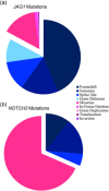
Incidence of all reported and novel JAG1 and NOTCH2 mutation types. (a) JAG1 protein‐truncating pathogenic variants are shown in blue color tones and include: frameshift (n = 303), nonsense (n = 113), splice site (n = 89), and gross deletion (n = 70). JAG1 non‐protein‐truncating pathogenic variants are shown in pink color tones and include: missense (n = 104), in‐frame deletion (n = 9), gross duplication (n = 3), translocation (n = 2), and inversion (n = 1). (b) NOTCH2 protein‐truncating pathogenic variants are shown in blue color tones and include: frameshift (n = 2), nonsense (n = 3), and splice site (n = 1). NOTCH2 non‐protein‐truncating pathogenic variants are shown in pink color tones and include: missense (n = 13)
Figure 3
Figure 3
JAG1 missense variants cluster in the N‐terminus. (a) Schematic showing all reported (lower) and novel (upper) missense mutations in JAG1. Dashed lines within the protein indicate exon boundaries and numbers indicate amino acid coordinates. (b) Distance in nucleotides between missense mutations within exons 1–6 and exons 7–26. Statistical significance (p < .0001) was calculated using an unpaired, two‐tailed t test. RefSeq NM_000214.2. Protein schematic was prepared using ProteinPaint software from Saint Jude Children's Research Hospital–Pediatric Cancer Data Portal (Zhou et al., 2016)
Figure 4
Figure 4
Gain of cysteine missense variants are more tolerated in JAG1. Schematic showing all missense variants involving gain (blue) or loss (red) of a cysteine in JAG1 from control samples present in gnomAD (lower) and in patients with ALGS (upper). The disease population includes combined data from HGMD, ClinVar, LOVD, and novel mutations reported here. Numbers within the circle indicate the number of alleles seen for each variant in gnomAD. Circle size and height is proportional to the number of probands with that variant in the disease population. The concentric circle in the ALGS cohort indicates multiple variants at the same amino acid position (p.C78Y, p.C78G, p.C78R, and p.C78S). RefSeq NM_000214.2. Protein schematic was prepared using ProteinPaint software from Saint Jude Children's Research Hospital–Pediatric Cancer Data Portal (Zhou et al., 2016)
Figure 5
Figure 5
Cysteine‐loss missense variants are defective in protein localization. Confocal microscopy of stably‐transfected NIH‐3T3 cells expressing the following controls: (a) wild type JAG1 and two positive controls with known nuclear retention and perinuclear localization (b) p.G274D and (c) p.L37S (Lu et al., 2003; Morrissette et al., 2001). (d–l) Cysteine‐loss missense variants all show protein clustering near the nuclei
Figure 6
Figure 6
Cysteine‐loss missense variants are differentially sensitive to enzymatic proteolysis. Western blot of protein lysates from stably transfected NIH‐3T3 cells treated with Endo H or Trypsin. Controls include wild type JAG1 (not sensitive), p.L37S (sensitive), and p.G274D (partially sensitive)
Figure 7
Figure 7
Luciferase assay of cysteine‐loss missense variants showing reduced JAG1 signaling. Luciferase assay of NIH‐3T3 cells transfected with 4XCBF‐luciferase reporter construct and cocultured with wild type or mutant JAG1‐expressing cells. RLU signals were normalized to internal Renilla controls. p.L37S and p.G274D are included as negative controls. All variants showed a statistically significant decrease in luciferase activity (unpaired, two‐tailed t test) when compared to wild type, with the exception of p.C693Y, which was statistically unchanged from wild type, and p.C714Y, which showed a statistically significant increase in luciferase activity from wild type. Red bars indicate variants that are not expressed on the cell membrane and blue bars indicate variants that have some partial expression on the cell membrane (based on results from Figures 5 and 6)

References

    1. Agrawal, S. , Chennuri, V. , & Agrawal, P. (2015). Genetic diagnosis in an Indian child with alagille syndrome. Indian Journal of Pediatrics, 82(7), 653–654. 10.1007/s12098-014-1684-0 - DOI - PubMed
    1. Alagille, D. , Estrada, A. , Hadchouel, M. , Gautler, M. , Odièvre, M. , & Dommergues, J. P. (1987). Syndromic paucity of interlobular bile ducts (Alagille syndrome or arteriohepatic dysplasia): Review of 80 cases. The Journal of Pediatrics, 110(2), 195–200. - PubMed
    1. Alagille, D. , Odièvre, M. , Gautier, M. , & Dommergues, J. P. (1975). Hepatic ductular hypoplasia associated with characteristic facies, vertebral malformations, retarded physical, mental, and sexual development, and cardiac murmur. The Journal of Pediatrics, 86(1), 63–71. - PubMed
    1. Andersson, E. R. , Chivukula, I. V. , Hankeova, S. , Sjöqvist, M. , Tsoi, Y. L. , Ramsköld, D. , … Lendahl, U. (2018). Mouse model of alagille syndrome and mechanisms of jagged1 missense mutations. Gastroenterology, 154(4), 1080–1095. 10.1053/j.gastro.2017.11.002 - DOI - PMC - PubMed
    1. Andrawes, M. B. , Xu, X. , Liu, H. , Ficarro, S. B. , Marto, J. A. , Aster, J. C. , & Blacklow, S. C. (2013). Intrinsic selectivity of notch 1 for delta‐like 4 over delta‐like 1. Journal of Biological Chemistry, 288(35), 25477–25489. 10.1074/jbc.M113.454850 - DOI - PMC - PubMed

Publication types