An important role of cutaneous lymphatic vessels in coordinating and promoting anagen hair follicle growth
- PMID: 31344105
- PMCID: PMC6657912
- DOI: 10.1371/journal.pone.0220341
An important role of cutaneous lymphatic vessels in coordinating and promoting anagen hair follicle growth
Abstract
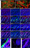
The lymphatic vascular system plays important roles in the control of tissue fluid homeostasis and immune responses. While VEGF-A-induced angiogenesis promotes hair follicle (HF) growth, the potential role of lymphatic vessels (LVs) in HF cycling has remained unknown. In this study, we found that LVs are localized in close proximity to the HF bulge area throughout the postnatal and depilation-induced hair cycle in mice and that a network of LVs directly connects the individual HFs. Increased LV density in the skin of K14-VEGF-C transgenic mice was associated with prolongation of anagen HF growth. Conversely, HF entry into the catagen phase was accelerated in K14-sVEGFR3 transgenic mice that lack cutaneous LVs. Importantly, repeated intradermal injections of VEGF-C promoted hair growth in mice. Conditioned media from lymphatic endothelial cells promoted human dermal papilla cell (DPC) growth and expression of IGF-1 and alkaline phosphatase, both activators of DPCs. Our results reveal an unexpected role of LVs in coordinating and promoting HF growth and identify potential new therapeutic strategies for hair loss-associated conditions.
Conflict of interest statement
The authors have declared that no competing interests exist.
Figures


Similar articles
-
HNG, A Humanin Analogue, Promotes Hair Growth by Inhibiting Anagen-to-Catagen Transition.Int J Mol Sci. 2020 Jun 26;21(12):4553. doi: 10.3390/ijms21124553. Int J Mol Sci. 2020. PMID: 32604799 Free PMC article.
-
Regulation of hair follicle development by exosomes derived from dermal papilla cells.Biochem Biophys Res Commun. 2018 Jun 2;500(2):325-332. doi: 10.1016/j.bbrc.2018.04.067. Epub 2018 Apr 13. Biochem Biophys Res Commun. 2018. PMID: 29654758
-
Thrombospondin-1 plays a critical role in the induction of hair follicle involution and vascular regression during the catagen phase.J Invest Dermatol. 2003 Jan;120(1):14-9. doi: 10.1046/j.1523-1747.2003.12045.x. J Invest Dermatol. 2003. PMID: 12535193
-
STAT5 Activation in the Dermal Papilla Is Important for Hair Follicle Growth Phase Induction.J Invest Dermatol. 2016 Sep;136(9):1781-1791. doi: 10.1016/j.jid.2016.04.014. Epub 2016 Apr 27. J Invest Dermatol. 2016. PMID: 27131881 Review.
-
Epithelial growth control by neurotrophins: leads and lessons from the hair follicle.Prog Brain Res. 2004;146:493-513. doi: 10.1016/S0079-6123(03)46031-7. Prog Brain Res. 2004. PMID: 14699982 Review.
Cited by
-
Advances in hair growth.Fac Rev. 2022 Jan 12;11:1. doi: 10.12703/r/11-1. eCollection 2022. Fac Rev. 2022. PMID: 35156098 Free PMC article. Review.
-
Tissue-resident macrophages specifically express Lactotransferrin and Vegfc during ear pinna regeneration in spiny mice.Dev Cell. 2024 Feb 26;59(4):496-516.e6. doi: 10.1016/j.devcel.2023.12.017. Epub 2024 Jan 15. Dev Cell. 2024. PMID: 38228141 Free PMC article.
-
Translational frontiers: insight from lymphatics in skin regeneration.Front Physiol. 2024 Feb 29;15:1347558. doi: 10.3389/fphys.2024.1347558. eCollection 2024. Front Physiol. 2024. PMID: 38487264 Free PMC article. Review.
-
Immune modulation of hair follicle regeneration.NPJ Regen Med. 2020 May 11;5:9. doi: 10.1038/s41536-020-0095-2. eCollection 2020. NPJ Regen Med. 2020. PMID: 32411394 Free PMC article. Review.
-
Dermal papilla cells-conditioned medium attenuates oxidative stress-induced senescence via ferroptosis inhibition.Sci Rep. 2025 Jul 9;15(1):24789. doi: 10.1038/s41598-025-08968-2. Sci Rep. 2025. PMID: 40634437 Free PMC article.
References
-
- Muller-Rover S, Handjiski B, van der Veen C, Eichmuller S, Foitzik K, McKay IA, et al. A comprehensive guide for the accurate classification of murine hair follicles in distinct hair cycle stages. J Invest Dermatol. 2001;117(1):3–15. Epub 2001/07/10. 10.1046/j.0022-202x.2001.01377.x . - DOI - PubMed
Publication types
MeSH terms
Substances
LinkOut - more resources
Full Text Sources
Other Literature Sources
Molecular Biology Databases
Research Materials
Miscellaneous

