. 2019 Jul 25;14(7):e0218726.
doi: 10.1371/journal.pone.0218726. eCollection 2019.

Dexmedetomidine-induced cardioprotection is mediated by inhibition of high mobility group box-1 and the cholinergic anti-inflammatory pathway in myocardial ischemia-reperfusion injury

Affiliations

Dexmedetomidine-induced cardioprotection is mediated by inhibition of high mobility group box-1 and the cholinergic anti-inflammatory pathway in myocardial ischemia-reperfusion injury

Juan Zhang et al. PLoS One. .

Abstract

Objectives: Dexmedetomidine (DEX) is a selective α2-adrenoceptor agonist that has anti-inflammatory and cardioprotective effects in myocardial ischemia/reperfusion (I/R) injury. The present study aimed to investigate the underlying mechanism by which DEX protects against myocardial I/R.

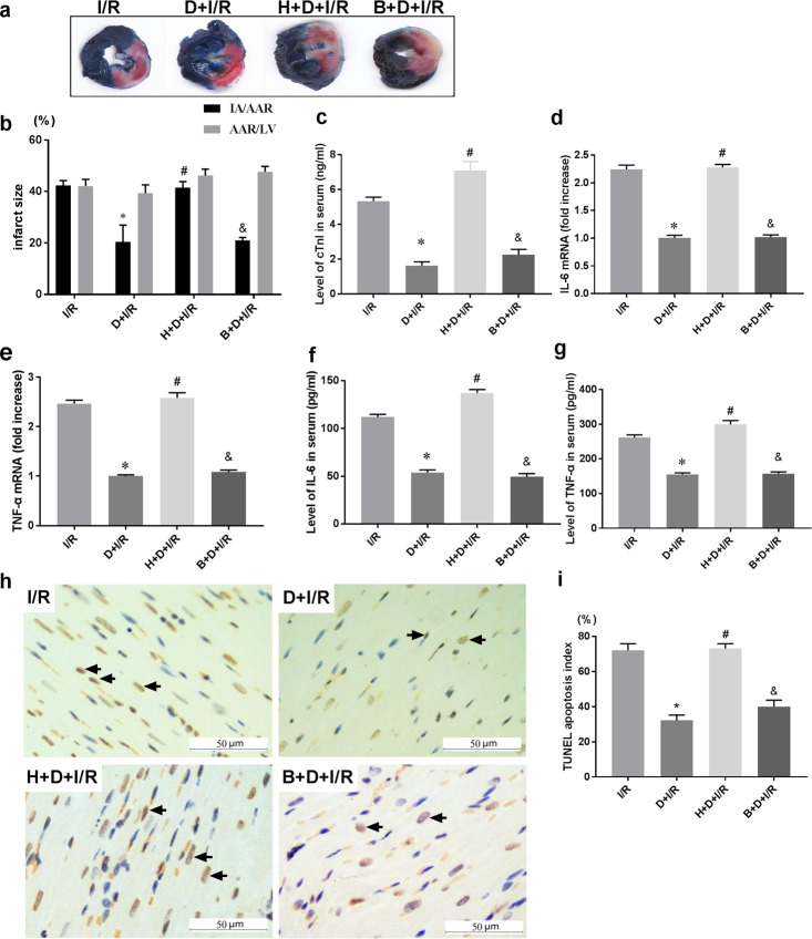
Methods: Sprague Dawley rats were subjected to either sham operation or myocardial I/R, which was induced by ligating the left anterior descending coronary artery for 30 min followed by reperfusion for 120 min. Rats were treated with either DEX or saline prior to surgery. We measured heart infarct size, serum cardiac Troponin I (cTnI), cardiac High mobility group box-1 (HMGB1) expression, myocardial apoptosis and cytokine production of interleukin-6 (IL-6) and tumor necrosis factor-α (TNF-α). Besides, we evaluated the heart function at 4 weeks post-reperfusion by echocardiography. Unilateral vagotomy or inhibition of the α7 nicotinic acetylcholine receptor (α7nAChR) with methyllycaconitine (MLA) was applied to investigate whether DEX-induced cardioprotection is mediated via the cholinergic anti-inflammatory pathway. Cardiac-selective overexpression of HMGB1 was administered to further confirm if HMGB1 is a key anti-inflammatory target during DEX-induced cardioprotection.

Results: DEX pretreatment significantly attenuated I/R-induced cardiac damage, as evidenced by decreases in short-term injury indicators including myocardial infarct size, cTnI release, myocardial apoptosis, cardiac HMGB1 expression, IL-6 and TNF-α production, as well as improvement on long-term cardiac function at 4 weeks post-reperfusion. These effects were partially reversed by either unilateral vagotomy or methyllycaconitine treatment. Besides, cardiac HMGB1-overexpression nearly abolished DEX-induced cardioprotection.

Conclusions: DEX pretreatment protects against myocardial I/R by inhibiting cardiac HMGB1 production and activating the cholinergic anti-inflammatory pathway.


Conflict of interest statement

The authors have declared that no competing interests exist.

Figures

Fig 1
Fig 1. Schematic diagram of experimental design.
Rat hearts were subjected to 30 min anterior descending coronary artery occlusion and 120 min of reperfusion. In protocol 1, unilateral vagotomy or sham operation was induced before DEX or saline pretreatment followed by myocardial I/R to determine the involvement of the vagal nerve. In protocol 2, MLA or saline was administered before DEX pretreatment to detect the involvement of the α7nAChR. In protocol 3, rats received an intrapericardial injection of AAV9-HMGB1 or AAV9-Blank and were subsequently subjected to either DEX or I/R treatment after 3 weeks. MLA: methyllycaconitine; AAV9: adeno-associated virus serotype 9; HMGB1: High mobility group box-1.
Fig 2
Fig 2. The vagal nerve mediates DEX-induced short-term cardioprotection against I/R.
Unilateral vagotomy or sham operation was induced before DEX or saline pretreatment followed by myocardial I/R. The extent of myocardial injury was evaluated by infarct size using Evans blue/TTC staining (a-b), serum levels of cTnI (c), and myocardial expression of IL-6 and TNF-α (d-e). Serum levels of IL-6 and TNF-α were also measured by ELISA (f-g). Myocardial apoptosis in infarct zone was evaluated by TUNEL staining (h). Representative pictures for each group as well as the corresponding quantitative analysis using the TUNEL apoptosis index (TUNEL-positive cell numbers /total cell numbers ×100%) were shown; arrows indicate positive cells, Scale bar = 50μm. Data shown are mean ± SEM; n = 10 per group; $ p<0.05 vs. S; *p < 0.05 vs. I/R; #p < 0.05 vs. D+I/R; &p < 0.05 vs. V+I/R. All specimen were obtained at the timepoint of myocardial I/R for 2hrs. S: sham operation; I/R: myocardial ischemia-reperfusion; D: dexmedetomidine; V: vagotomy;
Fig 3
Fig 3. The vagal nerve mediates DEX-induced long-term cardioprotection against I/R.
To investigate whether DEX exerts long-term cardioprotective effects and the involvement of the vagal nerve, echocardiography was performed to evaluate the LV function at 4 weeks post-I/R before animals were sacrificed. M-mode images at long-axis view were shown. Ejection fraction (EF) were calculated and compared among different groups: EF (%) = [(EDV−ESV)/EDV]×100%; EDV (end diastolic volume); ESV (end systolic volume). All measurements were based on the average of three consecutive cardiac cycles. Data shown are mean ± SEM; n = 6 per group; $ p<0.05 vs. S; *p < 0.05 vs. I/R; #p < 0.05 vs. D+I/R; &p < 0.05 vs. V+I/R. S: sham operation; I/R: myocardial ischemia-reperfusion; D: dexmedetomidine; V: vagotomy.;
Fig 4
Fig 4. The vagal nerve modulates DEX-induced downregulation of cardiac HMGB1 during I/R.
Unilateral vagotomy or sham operation was induced before DEX or saline pretreatment followed by myocardial I/R. HMGB1 protein levels in the left ventricles were measured using Western blot (a) and HMGB1 mRNA levels were assessed by qRT-PCR (b). Representative images of immunohistochemical staining for HMGB1 and quantitative results (c). Arrows indicate HMGB1-positive cells. Scale bar = 50μm. All specimen were obtained at the timepoint of myocardial I/R for 2hrs. Data shown are mean ± SEM; n = 6 per group; $ p<0.05 vs. S; *p < 0.05 vs. I/R; #p < 0.05 vs. D+I/R; &p < 0.05 vs. V+I/R. S: sham operation; I/R: myocardial ischemia-reperfusion; D: dexmedetomidine; V: vagotomy;
Fig 5
Fig 5. Blockade of the α7nAChR abolishes DEX-induced short-term cardioprotection against I/R.
Rats were subjected to intraperitoneal injection of saline or methyllycaconitine (MLA, α7nAChR antagonist) before DEX treatment. (a-b) Infarct sizes determined by Evans blue/TTC staining and representative images of each group are shown. (c) Serum cTnI levels; mRNA levels (d-e) and serum levels (f-g) of inflammatory cytokines including IL-6 and TNF-α were also detected. Myocardial apoptosis in infarct zone was evaluated by TUNEL staining (h) (arrows indicate positive cells, Scale bar = 50μm.); Data shown are mean ± SEM; n = 10 per group; $ p<0.05 vs. S; *p < 0.05 vs. I/R; #p < 0.05 vs. D+I/R; &p < 0.05 vs. M+I/R. All specimen were obtained at the timepoint of myocardial I/R for 2hrs. S: sham operation; I/R: myocardial ischemia-reperfusion; D: dexmedetomidine; M: methyllycaconitine.
Fig 6
Fig 6. Blockade of the α7nAChR abolishes DEX-induced long-term cardioprotection against I/R.
To investigate whether the α7nAChR was involved in the long-term cardioprotective effects of DEX, we administrate MLA, the selective antagonist of α7nAChR, prior to DEX treatment followed with I/R operation. After 4 weeks, the echocardiography was performed to evaluate the LV function before the animals were sacrificed. M-mode images at long-axis view were shown. Ejection fraction (EF) were calculated and compared among different groups: EF (%) = [(EDV−ESV)/EDV]×100%; EDV (end diastolic volume); ESV (end systolic volume). All measurements were based on the average of three consecutive cardiac cycles. Data shown are mean ± SEM; n = 6 per group; $ p<0.05 vs. S; *p < 0.05 vs. I/R; #p < 0.05 vs. D+I/R; &p < 0.05 vs. M+I/R. S: sham operation; I/R: myocardial ischemia-reperfusion; D: dexmedetomidine; M: methyllycaconitine.
Fig 7
Fig 7. Blockade of the α7nAChR reverses DEX-induced downregulation of HMGB1 during I/R.
To explore the involvement of α7nAChR, methyllycaconitine (MLA, α7nAChR antagonist) was administered. HMGB1 protein levels in the left ventricles were measured using Western blot (a) and HMGB1 mRNA levels were assessed by q-RT-PCR (b). Representative images of immunohistochemical staining for HMGB1 and quantitative results (c). Arrows indicate HMGB1-positive cells. Scale bar = 50 μm; n = 6 per group; $ p<0.05 vs. S; *p < 0.05 vs. I/R; #p < 0.05 vs. D+I/R; &p < 0.05 vs. M+I/R. S: sham operation; I/R: myocardial ischemia-reperfusion; D: dexmedetomidine; M: methyllycaconitine.
Fig 8
Fig 8. Cardiac-selective overexpression of HMGB1 using AAV9.
Intrapericardial injection of AAV9-HMGB1 (conjugated to green fluorescent protein) was performed to induce cardiac-selective HMGB1 overexpression; blank AAV9 (AAV9-Blank) was used as a control. Representative photographs of heart sections from both AAV9-HMGB1 and AAV9-Blank groups are shown in panel (a); mRNA levels of myocardial HMGB1 were quantified using qPCR (b). *p < 0.01 vs. AAV9-Blank.
Fig 9
Fig 9. Cardiac-selective HMGB1 overexpression eliminates DEX-induced short-term cardioprotection against I/R.
Rats were subjected to intrapericardial injection of AAV9-HMGB1 or AAV9-Blank as control 3 weeks before DEX pretreatment followed with the I/R operation. (a-b) Infarct size and representative images of each group are shown. (c) Serum cTnI levels; mRNA levels (d-e) and serum levels (f-g) of inflammatory cytokines including IL-6 and TNF-α were also detected. Myocardial apoptosis in infarct zone was evaluated by TUNEL staining (h-i) (arrows indicate positive cells, Scale bar = 50μm.); All specimen were obtained at the timepoint of myocardial I/R for 2hrs. Data shown are mean ± SEM; n = 10 per group; *p < 0.01 vs. I/R; #p < 0.05 vs. D+I/R; &p<0.05 vs. H+D+I/R; H: AAV9-HMGB1; B: AAV9-Blank; D: dexmedetomidine; I/R: myocardial ischemia-reperfusion;
Fig 10
Fig 10. Cardiac-selective HMGB1 overexpression eliminates DEX-induced long-term cardioprotection against I/R.
Rats were subjected to intrapericardial injection of AAV9-HMGB1 or AAV9-Blank as control before DEX pretreatment followed with the I/R operation. After another 4 weeks of reperfusion, the echocardiography was performed to evaluate the LV function before the animals were sacrificed. M-mode images at long-axis view were shown. Ejection fraction (EF) were calculated and compared among different groups: EF (%) = [(EDV−ESV)/EDV]×100%; EDV (end diastolic volume); ESV (end systolic volume). All measurements were based on the average of three consecutive cardiac cycles. Data shown are mean ± SEM; n = 6 per group; *p < 0.01 vs. I/R; #p < 0.05 vs. D+I/R; &p<0.05 vs. H+D+I/R; H: AAV9-HMGB1; B: AAV9-Blank; D: dexmedetomidine; I/R: myocardial ischemia-reperfusion;

References

    1. Finegold JA, Asaria P, Francis DP. Mortality from ischaemic heart disease by country, region, and age: statistics from World Health Organisation and United Nations. Int J Cardiol. 2013;168(2):934–45. 10.1016/j.ijcard.2012.10.046 - DOI - PMC - PubMed
    1. Ma A, Qi S, Chen H. Antioxidant therapy for prevention of inflammation, ischemic reperfusion injuries and allograft rejection. Cardiovasc Hematol Agents Med Chem. 2008;6(1):20–43. - PubMed
    1. Ando M, Katare RG, Kakinuma Y, Zhang D, Yamasaki F, Muramoto K, et al. Efferent vagal nerve stimulation protects heart against ischemia-induced arrhythmias by preserving connexin43 protein. Circulation. 2005;112(2):164–70. 10.1161/CIRCULATIONAHA.104.525493 - DOI - PubMed
    1. Wang Z, Yu L, Wang S, Huang B, Liao K, Saren G, et al. Chronic intermittent low-level transcutaneous electrical stimulation of auricular branch of vagus nerve improves left ventricular remodeling in conscious dogs with healed myocardial infarction. Circ Heart Fail. 2014;7(6):1014–21. 10.1161/CIRCHEARTFAILURE.114.001564 - DOI - PubMed
    1. Zhang J, Yong Y, Li X, Hu Y, Wang J, Wang YQ, et al. Vagal modulation of high mobility group box-1 protein mediates electroacupuncture-induced cardioprotection in ischemia-reperfusion injury. Sci Rep. 2015;5:15503 10.1038/srep15503 - DOI - PMC - PubMed

Publication types

MeSH terms