. 2019 Oct 15;20(20):2633-2642.
doi: 10.1002/cbic.201900299. Epub 2019 Aug 28.

Reconstitution and Coupling of DNA Replication and Segregation in a Biomimetic System

Affiliations

Reconstitution and Coupling of DNA Replication and Segregation in a Biomimetic System

Daniel Hürtgen et al. Chembiochem. .

Abstract

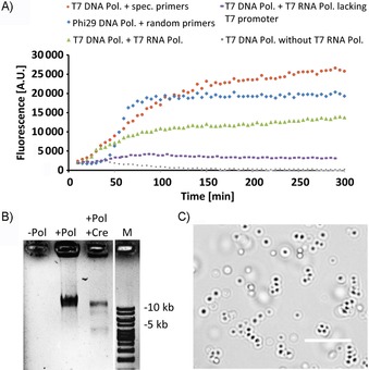
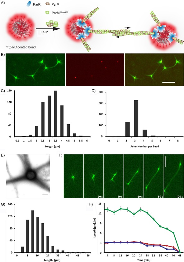
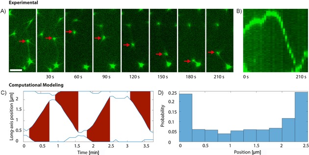
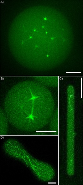
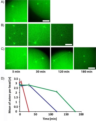
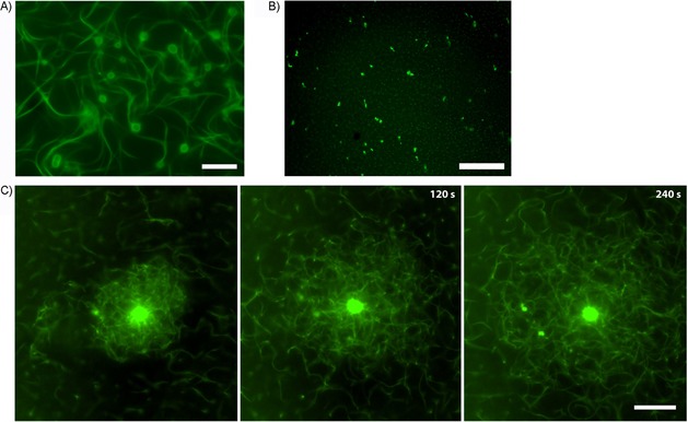
A biomimetic system capable of replication and segregation of genetic material constitutes an essential component for the future design of a minimal synthetic cell. Here we have used the simple T7 bacteriophage system and the plasmid-derived ParMRC system to establish in vitro DNA replication and DNA segregation, respectively. These processes were incorporated into biomimetic compartments providing an enclosed reaction space. The functional lifetime of the encapsulated segregation system could be prolonged by equipping it with ATP-regenerating and oxygen-scavenging systems. Finally, we showed that DNA replication and segregation processes could be coupled in vitro by using condensed DNA nanoparticles resulting from DNA replication. ParM spindles extended over tens of micrometers and could thus be used for segregation in compartments that are significantly longer than bacterial cell size. Overall, this work demonstrates the successful bottom-up assembly and coupling of molecular machines that mediate replication and segregation, thus providing an important step towards the development of a fully functional minimal cell.

Keywords: DNA nanoparticles; DNA replication; DNA segregation; ParM; T7; minimal cell.


Conflict of interest statement

The authors declare no conflict of interest.

Figures

Figure 1
Figure 1
Replication, recircularization, and compaction of a plasmid containing the T7 promoter. A) Time course of RCA‐based replication of pRepC plasmid containing the T7 promoter and loxP sites (depicted in Figure S1), measured as fluorescence of the DNA‐binding PicoGreen dye. Reaction mixtures contained, as indicated, Phi29 DNA polymerase, T7 DNA polymerase, and T7 RNA polymerase, as well as specific or random primers. For a control reaction with T7 DNA polymerase and T7 RNA polymerase, a pQE30 plasmid lacking the T7 promoter was used. B) Recircularization of the replicated plasmid, mediated by Cre recombinase. Where indicated, Cre recombinase was added after 16 h of replication, and the reaction mixture was incubated for another 30 min. Reaction mixtures were separated along with a DNA ladder (1 kb) on a Midori‐green stained agarose gel. The lower band migrating below 5 kb corresponds to the circularized DNA, whereas larger products apparently correspond to linear concatamers (Figure S2). C) DNA nanoparticles emerging upon prolonged (>12 h) T7 DNA replication reaction. Scale bar: 10 μm.
Figure 2
Figure 2
DNA segregation by the ParMRC system. A) Schematic representation of in vitro segregation of Cy3‐labeled parC sequences (red) bound to beads through streptavidin/biotin chemistry. Polymerization of Alexa488‐labeled ParM (green) leads to filament formation and segregation of beads in dependence on ParR (blue). B) In vitro reconstitution of the ParMRC system, with ParM (green) forming spindles connecting two beads, as well as free asters that are linked to the parC‐coated beads (red) by ParR (unlabeled). Reaction mixtures containing 5 μm ParM, 250 nm ParR, and 14 pm parC‐coated beads were mixed with 10 mm ATP to induce polymerization. Scale bar: 5 μm. C) Distribution of aster length, and D) number of asters per bead. E) TEM image of ParM filament bundles growing from a bead. Scale bar: 200 nm. F) Time‐lapse series of an elongating ParM spindle pushing parC‐bound beads apart. Scale bar: 10 μm. G) Distributions of spindle lengths. H) Stability of spindle length (green), aster length (red), and asters per bead (blue) over time.
Figure 3
Figure 3
DNA segregation as an ongoing, dynamic event. A) Time‐lapse imaging of one parC‐covered bead (red arrow) undergoing the segregation process multiple times between two neighboring beads within 210 s. Scale bar: 5 μm. B) A kymograph shows the localization dynamics of this bead. C) Simulation of ParMRC‐mediated bead segregation. Shown are the positions of three beads along the long axis of a rectangular compartment (blue lines). Red areas indicate spindle formation and extension. D) Distribution of bead positions along the long axis of the compartment resulting from simulation in (C), with approximately equal number of beads being positioned at each end of the compartment.
Figure 4
Figure 4
Segregation of parC‐coated beads in microcompartments. Several different types of biomimetic confinement were tested. A), B) Water‐in‐oil droplets. Scale bars: 10 μm and 5 μm, respectively. C) Water‐in‐oil droplet squeezed into PDMS channel. Scale bar: 20 μm. D) Half‐open PDMS channel covered with lipid bilayer isolated from E. coli. Scale bar: 10 μm.
Figure 5
Figure 5
Lifetime extension of the segregation system in water‐in‐oil droplets. Formation of ParM spindles at indicated time points for the system reconstituted A) as in Figure 2, B) with ATP regeneration, and C) with both ATP regeneration and an oxygen‐scavenger system. Scale bars: 5 μm. D) Quantification of aster formation in experiments shown in A (), B () and C (). Error bars indicate standard deviations.
Figure 6
Figure 6
Segregation of DNA nanoparticles by the ParMR system. A) DNA nanoparticles formed upon replication of the parC‐containing pRepC plasmid associate with Alexa488‐labeled ParM in a ParR‐dependent manner. Reaction conditions were as in Figure 2 F. B) Control reaction with pUC19 plasmid lacking parC. C) Time series of dynamic ParM filament meshwork formed around one large DNA nanoparticle that shears off and pushes apart smaller nanoparticles. Scale bars: 20 μm.

Similar articles

Cited by

References

    1. Göpfrich K., Platzman I., Spatz J. P., Trends Biotechnol. 2018, 36, 938. - PMC - PubMed
    1. Schwille P., Spatz J., Landfester K., Bodenschatz E., Herminghaus S., Sourjik V., Erb T. J., Bastiaens P., Lipowsky R., Hyman A., Dabrock P., Baret J.-C., Vidakovic-Koch T., Bieling P., Dimova R., Mutschler H., Robinson T., Tang T.-Y. D., Wegner S., Sundmacher K., Angew. Chem. Int. Ed. 2018, 57, 13382; - PubMed
    2. Angew. Chem. 2018, 130, 13566.
    1. Van Nies P., Westerlaken I., Blanken D., Salas M., Mencía M., Danelon C., Nat. Commun. 2018, 9, 1583. - PMC - PubMed
    1. Fischer H., Hinkle D. C., J. Biol. Chem. 1980, 255, 7956. - PubMed
    1. Schaerli Y., Stein V., Spiering M. M., Benkovic S. J., Abell C., Hollfelder F., Nucleic Acids Res. 2010, 38, e201. - PMC - PubMed

Publication types

MeSH terms

Substances

LinkOut - more resources