Review
. 2019 Jul 24;20(15):3622.
doi: 10.3390/ijms20153622.

Intervertebral Disc Nucleus Repair: Hype or Hope?

Affiliations
Review

Intervertebral Disc Nucleus Repair: Hype or Hope?

Gauri Tendulkar et al. Int J Mol Sci. .

Abstract

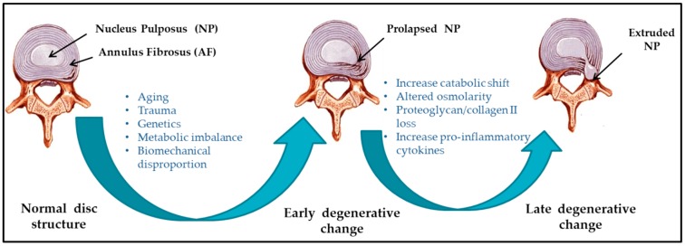
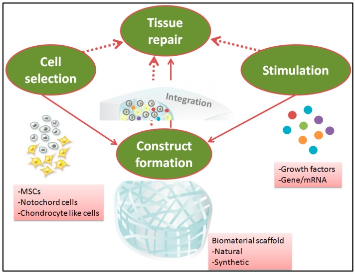
Chronic back pain is a common disability, which is often accredited to intervertebral disc degeneration. Gold standard interventions such as spinal fusion, which are mainly designed to mechanically seal the defect, frequently fail to restore the native biomechanics. Moreover, artificial implants have limited success as a repair strategy, as they do not alter the underlying disease and fail to promote tissue integration and subsequent native biomechanics. The reported high rates of spinal fusion and artificial disc implant failure have pushed intervertebral disc degeneration research in recent years towards repair strategies. Intervertebral disc repair utilizing principles of tissue engineering should theoretically be successful, overcoming the inadequacies of artificial implants. For instance, advances in the development of scaffolds aided with cells and growth factors have opened up new possibilities for repair strategies. However, none has reached the stage of clinical trials in humans. In this review, we describe the hitches encountered in the musculoskeletal field and summarize recent advances in designing tissue-engineered constructs for promoting nucleus pulposus repair. Additionally, the review focuses on the effect of biomaterial aided with cells and growth factors on achieving effective functional reparative potency, highlighting the ways to enhance the efficacy of these treatments.

Keywords: intervertebral disc; nucleus pulposus; repair; replacement; scaffold.


Conflict of interest statement

The authors declare no conflict of interest.

Figures

Figure 1
Figure 1
Schematic illustration of intervertebral disc (IVD) pathophysiology during degeneration.
Figure 2
Figure 2
Nucleus replacement devices. (A) The Aquerelle poly (vinyl alcohol) hydrogel has a swelling pressure similar to the nucleus pulposus (NP) in vivo. Once implanted, its final volume depends on the water content at equilibrium (reprinted with permission from Stryker Spine, Allendale, NJ, USA). (B) The Prosthetic disc nucleus (PDN-SOLO) device in dehydrated (left) and hydrated (right; as indicated by arrow) states. This device was designed to swell both in height and width within the disc space. The porous polyethylene weave allows fluid to pass into the hydrophilic core, which causes the device to expand vertically and horizontally. This process maximizes the device’s footprint on the vertebral endplates (reprinted with permission from Raymedica Inc., Minneapolis, MN, USA). (C) The Neudisc hydrogel, pre-hydration (left) and post-hydration (right). Hydration occurs in an anisotropic fashion, mainly in the vertical plane (reprinted with permission from Replication Medical, Inc., New Brunswick, NJ, USA). (D) The Newcleus Spiral Implant; once implanted, the device reconstitutes its original spiral shape. It localizes in place of the nucleus pulposus, which reconstitutes the volume, sparing the annular fibers (reprinted with permission from Zimmer Spine, Warsaw, IN, USA) [10,45].
Figure 3
Figure 3
Synergetic tissue engineering strategies for nucleus pulposus repair.

Similar articles

Cited by

References

    1. Clouet J., Fusellier M., Camus A., Le Visage C., Guicheux J. Intervertebral disc regeneration: From cell therapy to the development of novel bioinspired endogenous repair strategies. Adv. Drug Deliv. Rev. 2018 doi: 10.1016/j.addr.2018.04.017. - DOI - PubMed
    1. Iatridis J.C., Kang J., Kandel R., Risbud M.V. New horizons in spine research: Intervertebral disc repair and regeneration. J. Orthop. Res. 2017;35:5–7. doi: 10.1002/jor.23499. - DOI - PMC - PubMed
    1. Ahsan R., Tajima N., Chosa E., Sugamata M., Sumida M., Hamada M. Biochemical and morphological changes in herniated human intervertebral disc. J. Orthop. Sci. 2001;6:510–518. doi: 10.1007/s007760100006. - DOI - PubMed
    1. Buckwalter J.A. Aging and degeneration of the human intervertebral disc. Spine. 1995;20:1307–1314. doi: 10.1097/00007632-199506000-00022. - DOI - PubMed
    1. Ding F., Shao Z.W., Xiong L.M. Cell death in intervertebral disc degeneration. Apoptosis. 2013;18:777–785. doi: 10.1007/s10495-013-0839-1. - DOI - PubMed

MeSH terms

Substances