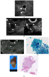
Endoscopic Ultrasound for Early Diagnosis of Pancreatic Cancer
- PMID: 31344904
- PMCID: PMC6787710
- DOI: 10.3390/diagnostics9030081
Endoscopic Ultrasound for Early Diagnosis of Pancreatic Cancer
Abstract
Detection of small pancreatic cancers, which have a better prognosis than large cancers, is needed to reduce high mortality rates. Endoscopic ultrasound (EUS) is the most sensitive imaging modality for detecting pancreatic lesions. The high resolution of EUS makes it particularly useful for detecting small pancreatic lesions that may be missed by other imaging modalities. Therefore, EUS should be performed in patients with obstructive jaundice in whom computed tomography (CT) or magnetic resonance imaging (MRI) does not identify a definite pancreatic lesion. Interest in the use of EUS for screening individuals at high risk of pancreatic cancer, including those with intraductal papillary mucinous neoplasms (IPMNs) and familial pancreatic cancer is growing. Contrast-enhanced EUS can facilitate differential diagnosis of small solid pancreatic lesions as well as malignant cystic lesions. In addition, EUS-guided fine needle aspiration can provide samples of small pancreatic lesions. Thus, EUS and EUS-related techniques are essential for early diagnosis of pancreatic cancer.
Keywords: contrast-enhanced endoscopic ultrasound; endoscopic ultrasound; pancreatic cancer.
Conflict of interest statement
M.K. received a speaker’s fee from the Olympus Corporation. The other authors declare no conflicts of interest relevant to this article.
Figures

Similar articles
-
Endoscopic ultrasonography for pancreatic solid lesions.J Med Ultrason (2001). 2020 Jul;47(3):377-387. doi: 10.1007/s10396-019-00959-x. Epub 2019 Aug 6. J Med Ultrason (2001). 2020. PMID: 31385143 Review.
-
Role of endoscopic ultrasound in the screening and follow-up of high-risk individuals for familial pancreatic cancer.World J Gastroenterol. 2019 Sep 14;25(34):5082-5096. doi: 10.3748/wjg.v25.i34.5082. World J Gastroenterol. 2019. PMID: 31558858 Free PMC article. Review.
-
Endoscopic diagnosis of cystic lesions of the pancreas.Dig Endosc. 2019 Jan;31(1):5-15. doi: 10.1111/den.13257. Epub 2018 Sep 30. Dig Endosc. 2019. PMID: 30085364 Review.
-
Conventional versus contrast-enhanced harmonic endoscopic ultrasonography-guided fine-needle aspiration for diagnosis of solid pancreatic lesions: A prospective randomized trial.Pancreatology. 2015 Sep-Oct;15(5):538-541. doi: 10.1016/j.pan.2015.06.005. Epub 2015 Jun 24. Pancreatology. 2015. PMID: 26145837 Clinical Trial.
-
High risk of acute pancreatitis after endoscopic ultrasound-guided fine needle aspiration of side branch intraductal papillary mucinous neoplasms.Endosc Ultrasound. 2015 Apr-Jun;4(2):109-14. doi: 10.4103/2303-9027.156728. Endosc Ultrasound. 2015. PMID: 26020044 Free PMC article.
Cited by
-
Screening of pancreatic cancer: Target population, optimal timing and how?Ann Med Surg (Lond). 2022 Nov 5;84:104814. doi: 10.1016/j.amsu.2022.104814. eCollection 2022 Dec. Ann Med Surg (Lond). 2022. PMID: 36582884 Free PMC article. Review.
-
Basic Principles and Role of Endoscopic Ultrasound in Diagnosis and Differentiation of Pancreatic Cancer from Other Pancreatic Lesions: A Comprehensive Review of Endoscopic Ultrasound for Pancreatic Cancer.J Clin Med. 2024 Apr 28;13(9):2599. doi: 10.3390/jcm13092599. J Clin Med. 2024. PMID: 38731128 Free PMC article. Review.
-
Pancreatic duct imaging during aging.Endosc Ultrasound. 2023 Mar-Apr;12(2):200-212. doi: 10.4103/EUS-D-22-00119. Endosc Ultrasound. 2023. PMID: 37148134 Free PMC article. Review.
-
Impact of circulating tumor DNA in hepatocellular and pancreatic carcinomas.J Cancer Res Clin Oncol. 2020 Jul;146(7):1625-1645. doi: 10.1007/s00432-020-03219-5. Epub 2020 Apr 27. J Cancer Res Clin Oncol. 2020. PMID: 32338295 Free PMC article. Review.
-
An optimal curriculum for training in endoscopic ultrasound: a summarized evidence-based literature systematic review.Surg Endosc. 2025 Jul;39(7):4076-4093. doi: 10.1007/s00464-025-11783-5. Epub 2025 May 23. Surg Endosc. 2025. PMID: 40410620 Review.
References
-
- The Editorial Board of the Cancer Statistics in Japan . Foundation for Promotion of Cancer Research (FPCR); 2017. [(accessed on 1 June 2019)]. Cancer Registry and Statistics. Cancer Information Service NCCJ (2018) Cancer Statistics in Japan. Available online: https://ganjoho.jp/en/professional/statistics/brochure/2017_en.html?
-
- Noone A.M., Howlader N., Krapcho M., Miller D., Brest A., Yu M., Ruhl J., Tatalovich Z., Mariotto A., Lewis D.R., et al. SEER Cancer Statistics Review, 1975–2015. [(accessed on 1 June 2019)];2018 Published. Available online: https://seer.cancer.gov/csr/1975_2015/
Publication types
LinkOut - more resources
Full Text Sources

