. 2019 Jul 24;24(15):2692.
doi: 10.3390/molecules24152692.

Anti-Biofilm Property of Bioactive Upconversion Nanocomposites Containing Chlorin e6 against Periodontal Pathogens

Affiliations

Anti-Biofilm Property of Bioactive Upconversion Nanocomposites Containing Chlorin e6 against Periodontal Pathogens

Tianshou Zhang et al. Molecules. .

Abstract

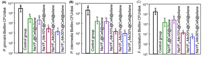
Photodynamic therapy (PDT) based periodontal disease treatment has received extensive attention. However, the deep tissue location of periodontal plaque makes the conventional PDT encounter a bottleneck. Herein, upconversion fluorescent nanomaterial with near-infrared light excitation was introduced into the treatment of periodontal disease, overcoming the limited tissue penetration depth of visible light in PDT. Photosensitizer Ce6 molecules were combined with upconversion nanoparticles (UCNPs) NaYF4:Yb,Er with a novel strategy. The hydrophobic UCNPs were modified with amphiphilic silane, utilizing the hydrophobic chain of the silane to bind to the hydrophobic groups of the UCNPs through a hydrophobic-hydrophobic interaction, and the Ce6 molecules were loaded in this hydrophobic layer. This achieves both the conversion of the hydrophobic to the hydrophilic surface and the loading of the oily photosensitizer molecules. Because the excitation position of the Ce6 molecule is in the red region, Mn ions were doped to enhance red light, and thus the improved PDT function. This Ce6 loaded UCNPs composites with efficient red upconversion luminescence show remarkable bacteriological therapeutic effect on Porphyromonas gingivalis, Prevotella intermedia and Fusobacterium nucleatum and the corresponding biofilms under 980 nm irradiation, indicating a high application prospect in the treatment of periodontal diseases.

Keywords: antibacterial photodynamic therapy; manganese ion; periodontitis; photosensitizer; upconversion nanoparticles.


Conflict of interest statement

The authors declare no conflict of interest. The funders had no role in the design of the study; in the collection, analyses, or interpretation of data; in the writing of the manuscript, or in the decision to publish the results.

Figures

Figure 1
Figure 1
The schematic diagram of the composite structure.Ce6 and NaYF4:Yb,Er UCNPs were combined with amphiphilic silane modification through hydrophobic-hydrophobic interaction. Hydrophobic side chain of the silane (blue) and hydrophobic group (green) on the surface of NaYF4:Yb,Er UCNPs can attach each other forming the close coating layer. After entering into the bacteria in the area of periodontal disease, singlet oxygen (1O2) can be effectively produced under the irradiation of 980 nm irradiation as shown in the figure. Mn doping greatly enhances the red emission and then the aPDT function.
Figure 2
Figure 2
Characterization of the NaYF4:Yb3+,Er3+,Mn2+ (0%, 10%, 20%, 30%)UCNPs. (A) and (B) are the TEM images of NaYF4:Yb3+,Er3+ and silane modified NaYF4:Yb3+,Er3+UCNPs, respectively. Color insets in (A,B) showed the schematic structures of nanoparticles. HR-TEM insets in B showed the high magnification of single nanoparticles. (C) FT-IR spectra of NaYF4:Yb3+,Er3+ and silane modified NaYF4:Yb3+,Er3+UCNPs. (D) XRD pattern of NaYF4:Yb3+,Er3+,Mn2+ (0%, 10%, 20%, 30%) UCNPs. The inset is the amplified diffraction peak of 111 plane of the NaYF4:Yb3+,Er3+,Mn2+ (10%, 20%, 30%) UCNPs. (E) Upconversion fluorescence spectra of NaYF4:Yb3+,Er3+,Mn2+ (0%, 10%, 20%, 30%) UCNPs with 980 nm excitation. The inset is the lifetime of the 2H11/2 energy level corresponding to green emission. (F) Proposed energy transfer mechanisms under the excitation of 980 nm NIR.
Figure 3
Figure 3
(A) Absorption spectrum and the upconversion spectrum of NaYF4:Yb3+,Er3+@Ce6@silane. Red emission region overlaps the absorption band of Ce6 molecules. (B) Generation of singlet oxygen generation under 980 nm irradiation. (C) Dark cytotoxicity of NaYF4:Yb3+,Er3+@Ce6@silane NPs on mouse fibroblast cell line L929: Viability of L929 cells vs. concentration of NaYF4:Yb3+,Er3+@Ce6@silane NPs after 24 h without irradiation in the dark (mean ± sd).
Figure 4
Figure 4
Images of live/dead cells of 4-day biofilms of three of P. gingivalis (left column), P. intermedia (middle column) and F. nucleatum (right column) on dentin squares for (AC) NIR control, (DF) NIR + NaYF4@Ce6@silane, (GI) NIR + NaYF4-Mn10%@Ce6@silane, (JL) NIR + NaYF4-Mn20%@Ce6@silane, and (MO) NIR + NaYF4-Mn30%@Ce6@silane. All images had the same scale bar as shown in (A). Live bacteria were stained green. Bacteria with compromised membranes were stained red. Live and dead bacteria together or on the top of each other yielded yellow/orange colors. UCNPs with increasing Mn doping content showed more red staining of compromised bacteria under NIR (980 nm) irradiation.
Figure 5
Figure 5
CFU counts of 4-day biofilms of (A) P. gingivalis, (B) P. intermedia and (C) F. nucleatum after PDT application (mean ± sd; n = 6). Nanoparticles with Mn doping would significantly decrease the CFU counts of all three-species biofilms. Note the log scale for the y-axis. Dissimilar letters indicated significant differences between each group (p < 0.05).
Figure 6
Figure 6
Polysaccharide production by 4-day biofilms of (A) P. gingivalis, (B) P. intermedia and (C) F. nucleatum on dentin squares (mean ± sd; n = 6). Dissimilar letters indicated significant differences between each group (p < 0.05).

References

    1. Ravald N., Johansson C.S. Tooth loss in periodontally treated patients. A long-term study of periodontal disease and root caries. J. Clin. Periodontol. 2012;39:73–79. doi: 10.1111/j.1600-051X.2011.01811.x. - DOI - PubMed
    1. Umeda M., Takeuchi Y., Noguchi K., Huang Y., Koshy G., Ishikawa I. Effects of nonsurgical periodontal therapy on the microbiota. Periodontolol. 2000. 2004;36:98–120. doi: 10.1111/j.1600-0757.2004.03675.x. - DOI - PubMed
    1. Pfitzner A., Sigusch B.W., Albrecht V., Glockmann E. Killing of periodontopathogenic bacteria by photodynamic therapy. J. Periodontol. 2004;75:1343–1349. doi: 10.1902/jop.2004.75.10.1343. - DOI - PubMed
    1. Zhu H., Fang Y., Miao Q., Qi X., Ding D., Chen P., Pu K. Regulating near-infrared photodynamic properties of semiconducting polymer nanotheranostics for optimized cancer therapy. ACS Nano. 2017;11:8998–9009. doi: 10.1021/acsnano.7b03507. - DOI - PubMed
    1. Carrera E., Dias H., Corbi S., Marcantonio R., Bernardi A., Bagnato V.S., Hamblin M., Rastelli A.N.d.S. The application of antimicrobial photodynamic therapy, (aPDT) in dentistry: a critical review. Laser Phys. 2016 doi: 10.1088/1054-660X/26/12/123001. - DOI - PMC - PubMed

LinkOut - more resources