. 2019 Apr;31(5):192-202.
doi: 10.1080/08958378.2019.1643955. Epub 2019 Jul 26.

Inhalation exposure to multi-walled carbon nanotubes alters the pulmonary allergic response of mice to house dust mite allergen

Affiliations

Inhalation exposure to multi-walled carbon nanotubes alters the pulmonary allergic response of mice to house dust mite allergen

Mark D Ihrie et al. Inhal Toxicol. 2019 Apr.

Abstract

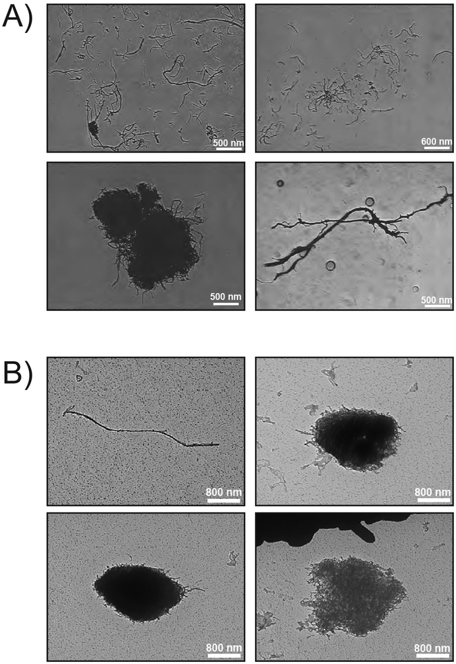
Background: Increasing evidence from rodent studies indicates that inhaled multi-walled carbon nanotubes (MWCNTs) have harmful effects on the lungs. In this study, we examined the effects of inhalation exposure to MWCNTs on allergen-induced airway inflammation and fibrosis. We hypothesized that inhalation pre-exposure to MWCNTs would render mice susceptible to developing allergic lung disease induced by house dust mite (HDM) allergen. Methods: Male B6C3F1/N mice were exposed by whole-body inhalation for 6 h a day, 5 d a week, for 30 d to air control or 0.06, 0.2, and 0.6 mg/m3 of MWCNTs. The exposure atmospheres were agglomerates (1.4-1.8 µm) composed of MWCNTs (average diameter 16 nm; average length 2.4 µm; 0.52% Ni). Mice then received 25 µg of HDM extract by intranasal instillation 6 times over 3 weeks. Necropsy was performed at 3 and 30 d after the final HDM dose to collect serum, bronchoalveolar lavage fluid (BALF), and lung tissue for histopathology. Results: MWCNT exposure at the highest dose inhibited HDM-induced serum IgE levels, IL-13 protein levels in BALF, and airway mucus production. However, perivascular and peribronchiolar inflammatory lesions were observed in the lungs of mice at 3 d with MWCNT and HDM, but not MWCNT or HDM alone. Moreover, combined HDM and MWCNT exposure increased airway fibrosis in the lungs of mice. Conclusions: Inhalation pre-exposure to MWCNTs inhibited HDM-induced TH2 immune responses, yet this combined exposure resulted in vascular inflammation and airway fibrosis, indicating that MWCNT pre-exposure alters the immune response to allergens.

Keywords: Carbon nanotubes; asthma; atopy; house dust mite allergen; inhalation; nanoparticles.


Conflict of interest statement

Declaration of Interests

The authors do not have any conflict of interest to report.

Figures

Fig. 1.
Fig. 1.
Representative photomicrographs of MWCNTs. A) TEM images depicting microscale morphometry of bulk MWCNTs, both entangled (bottom left) and unentangled (bottom right and top). B) TEM images depicting microscale morphometry of MWCNTs collected from the 0.06 mg/m3 chamber; entangled (top right and bottom) and unentangled (top left).
Fig. 2.
Fig. 2.
Illustration of exposure protocol. Mice were pre-exposed to MWCNTs (0.06, 0.2 or 0.6 mg/m3) by whole body inhalation for 30 days (5 days/week, 6 hr/day). After a 1 week acclimation period, the animals were sensitized with HDM extract (25 μg) via intranasal aspiration (days 0, 2, 4) and challenged with HDM extract (days 14, 16, 18). Mice were euthanized and necropsy performed on day 21 (3 days post-HDM) or 48 (30 days post-HDM).
Fig. 3.
Fig. 3.
MWCNT inclusions in alveolar macrophages. A) Representative images of alveolar macrophages from the 0.6 mg/m3 exposure group at 3 days post-HDM vehicle isolated from BALF by Cytospin centrifugation (left panel 200x, right panel 1000x). B) Numbers of macrophages with MWCNT inclusions per 500 cells at 3 days. C) Images of macrophages from the 0.6 mg/m3 exposure group at 30 days post-HDM vehicle (left panel 200x, right panel 1000x). D) Numbers of macrophages with MWCNT inclusions per 500 cells at 30 days.
Fig. 4.
Fig. 4.
MWCNT inclusions in alveolar macrophages in situ. MWCNTs were observed in alveolar macrophages in Alcian blue/periodic acid-Schiff stained lung sections at both 3 days (A) and 30 days post-HDM treatment (B). M1–M3 refers to images from 3 individual mice out of a total of 5 per group. Clusters of macrophages with MWCNT inclusions in alveolar spaces were seen only in the lungs of HDM-treated mice (see M3 in panel A).
Fig. 5.
Fig. 5.
Differential cell counts in BALF from mice at 3 or 30 days post-HDM treatment following inhalation pre-exposure to MWCNTs. A) Eosinophil numbers were elevated by HDM treatment with or without MWCNT inhalation pre-exposure. B) Neutrophils numbers marginally increased at the lowest dose of MWCNTs at 3 or 30 days. C) Numbers of alveolar macrophages remained largely unchanged at 3 or 30 days post-HDM treatment.
Fig. 6.
Fig. 6.
Serum IgE and BALF levels of IL-13 measured by ELISA at 3 days post-HDM treatment. A) HDM significantly enhanced IgE levels over corresponding controls, while MWCNT pre-exposure decreased IgE levels at the highest dose (0.6 μg/m3). *p<0.05 vs. vehicle, **p<0.01 vs. vehicle, #p<0.05 vs 0 mg/m3, ##p<0.01 vs. 0 mg/m3, ^p<0.05 vs. 0 mg/m3+HDM. B) IL-13 in BALF was suppressed by MWCNT pre-exposure (0.2 μg/m3). #p<0.05 vs 0 mg/m3, ^p<0.05 vs. 0 mg/m3+HDM.
Fig. 7.
Fig. 7.
Alcian Blue/Periodic Acid-Schiff staining for airway mucus. A) Representative AB-PAS-stained lung sections at 3 days post-HDM treatment. HDM treatment caused induced airway mucus production (arrows), while MWCNT pre-exposure at the highest dose blunted HDM-induced AB-PAS staining. B) Quantification of the AB-PAS+ area of mucosubstances normalized to airway length (units for area and length are arbitrary pixels). *p<0.05, HDM alone compared to HDM + MWCNT (0.6 mg/m3) as determined by Student’s t-test.
Fig. 8.
Fig. 8.
Representative hematoxylin and eosin stained lung sections at 3 days post-HDM treatment. Inflammatory lesions were observed around the terminal bronchioles (TB) and vessels in the lungs of mice treated with MWCNTs and HDM as indicated by the arrows. Images were captured at 100x and 400x.
Fig. 9.
Fig. 9.
Levels of pro-fibrotic cytokines in BALF measured by ELISA at 30 days post-HDM treatment. A) IL-1β was increased at 30 days by the highest dose of MWCNT (**p<0.01 vs 0 mg/m3). B) TGF-β1 was increased at 30 days and showed an increasing trend with MWCNT exposure (*p<0.05 vs 0 mg/m3).
Fig. 10.
Fig. 10.
Assessment of airway fibrosis at 30 days post-HDM exposure. A) Representative images of lung sections stained with Gomori’s trichrome showing blue collagen deposition around airways and blood vessels (arrows). B) Semi-quantitative morphometry of collagen thickness around airways larger than 300 × 300 μm accomplished using an area/perimeter ratio method. *p<0.05 vs 0.6 mg/m3 MWCNT + HDM.

References

    1. Akinbami LJ, Moorman JE, Bailey C, Zahran HS, King M, Johnson C. a, and Liu X (2012). Trends in asthma prevalence, health care use, and mortality in the United States, 2001–2010. NCHS Data Brief, 1–8. - PubMed
    1. Baughman RH, Zakhidov AA, and de Heer WA (2002). Carbon Nanotubes--the Route Toward Applications. Science, 297, 787–792. - PubMed
    1. Bonner JC (2010). Nanoparticles as a potential cause of pleural and interstitial lung disease. Proc. Am. Thorac. Soc, 7, 138–141. - PMC - PubMed
    1. Bonner JC, Silva RM, Taylor AJ, Brown JM, Hilderbrand SC, Castranova V, Porter D, Elder A, Oberdörster G, Harkema JR, Bramble LA, Kavanagh TJ, Botta D, Nel A, and Pinkerton KE (2013). Interlaboratory Evaluation of Rodent Pulmonary Responses to Engineered Nanomaterials: The NIEHS Nano GO Consortium. Environ. Health Perspect, 121, 676–682. - PMC - PubMed
    1. Dahm MM, Schubauer-Berigan MK, Evans DE, Birch ME, Bertke S, Beard JD, Erdely A, Fernback JE, Mercer RR, Grinshpun SA. (2018) Exposure assessments for a cross-sectional epidemiologic study of US carbon nanotube and nanofiber workers. Int J Hyg Environ Health. 221:429–440. - PubMed

Publication types

LinkOut - more resources