

Hemoptysis secondary to rupture of infected aortic aneurysm- a case report
- PMID: 31345241
- PMCID: PMC6659196
- DOI: 10.1186/s13019-019-0959-y
Hemoptysis secondary to rupture of infected aortic aneurysm- a case report
Abstract
Background: Massive hemoptysis is a life-threatening condition and can arise as a complication of various conditions. It rarely occurs as a complication of a ruptured thoracic aortic aneurysm. Even rarer are conditions where pseudoanurysms of aorta result due to infection.
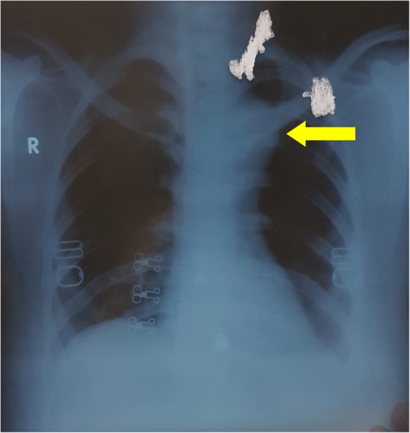
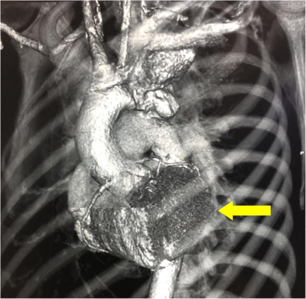
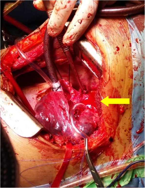
Case presentation: A 30 year-old female patient presented with left sided chest pain, intermittent fever, cough and massive hemoptysis. A pseudo-aneurysm of proximal descending thoracic aorta at the level of the left Subclavian artery was noted over CT scan. Upon performing a left posterolateral thoracotomy, the aneurysm was seen to have ruptured into the apical segment of left upper lobe, contained mainly by a thrombus. The anterior wall of the pseudoaneurysm was debrided and a bovine pericardial patch was used to repair the aortic defect. Cultures of the tissue obtained showed Enterobacter species, therefore the patient was prescribed 6 weeks of IV antibiotics following surgery. Post-operative CT scan revealed reduced diameter of the aorta. She was discharged in good health and remains well at follow up evaluation.
Conclusions: We present a case of hemoptysis caused by a ruptured descending aorta aneurysm into left lung. The aneurysm was secondary to infection by Enterobacter. Surgical repair of the concerned region of aorta was effective, without any major sequelae. To the best of our knowledge, no such cases have been reported previously.
Keywords: Aortic aneurysm; Hemoptysis; Infection; Rupture.
Conflict of interest statement
The authors declare that they have no competing interests.
Figures
References
-
- Coselli JS, Bosinovski J, LeMaire SA. Arch aneurysms in mastery of cardiothoracic surgery, vol. 2008. 2nd ed. Philadelphia: Lippincott Williams Wilkins. p. 556–8.
-
- Phang LY, Wong D, Agasthian T. Management of life threatening hemoptysis. Asian Cardiovasc Thorac Ann. 2001;9:200–203. doi: 10.1177/021849230100900309. - DOI
Publication types
MeSH terms
LinkOut - more resources
Full Text Sources
