. 2019 Jul 25;19(1):53.
doi: 10.1186/s40644-019-0238-0.

Imaging and clinical features of Castleman Disease

Affiliations

Imaging and clinical features of Castleman Disease

Shuang Zhao et al. Cancer Imaging. .

Abstract

Background: Castleman disease (CD) is a group of uncommon lymphoproliferative disorders that is easily confused with lymphoma or other solid tumors. The purpose of our study was to evaluate the imaging and clinical findings of CD, and thus improve the understanding and diagnosis of CD.

Methods: This retrospective study included 74 patients (37 men and 37 women, mean age ± standard deviation, 35 ± 15.2 years,) with histopathologically confirmed CD diagnosed based on CT or MRI between January 2010 and May 2017. The CT and MRI findings were analyzed by two radiologists in consensus, and clinical presentation and histopathologic characteristics were documented.

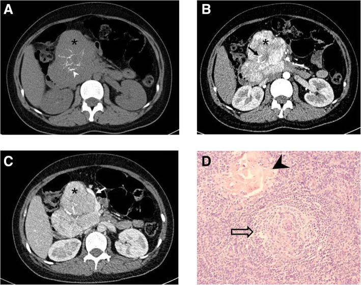
Results: The CD subtypes included 61 hyaline vascular variant cases (82.4%) and 13 plasma cell variant cases (17.6%). Unicentric CD and multicentric CD were observed in 65 (87.8%) and 9 (12.2%) patients, respectively. On non-enhanced CT, enlarged nodes with hypodensity or isodensity were seen, whereas varying degrees of enhancement were observed in contrast-enhanced CT. Homogeneous and heterogeneous enhancements were observed in 43 (62.3%) and 26 (37.7%) patients, respectively. Hypertrophied vessels and calcification were detected in 38 (51.2%) and 18 (24.3%) patients, respectively. MRI revealed hypointense to isointense lesions on T1-weighted images, isointense to hyperintense lesions on T2-weighted images, and hyperintense lesions on diffusion-weighted imaging; 9 (75%) and 3 (25%) patients demonstrated homogeneous and heterogeneous enhancement, respectively.

Conclusion: CD often shows well-defined, mildly hypodense or isodense, homogeneous lymph nodules on non-enhanced CT/MRI, with intermediate and marked enhancement on contrast-enhanced CT/MRI. Calcification and hypertrophied vessels may be valuable diagnostic features.

Keywords: Castleman disease; Computed tomography; Diffusion-weighted imaging; Magnetic resonance imaging.


Conflict of interest statement

The authors declare that they have no competing interests.

Figures

Fig. 1
Fig. 1
HVV unicentric Castleman disease in a 26-year-old woman. Axial non-enhanced (a), arterial phase (b), and portal venous phase (c) computed tomography images of the abdomen depict a well-defined heterogeneous mass of soft-tissue density (*) with branched calcification at the pancreatic head (arrowhead), which shows progressive enhancement, slightly greater than that of the pancreas. The feeding artery (black arrow) and draining vein (white arrow) can be seen. Photomicrograph (d, original magnification, × 200; hematoxylin-eosin [H-E] staining) shows marked vascular proliferation and hyalinization of the abnormal germinal center, with a tight concentric layering of lymphocytes at the periphery of the follicle, resulting in an “onion-skin” appearance (open arrow) and vessel-rich interfollicular stroma (arrowhead). (UCD = unicentric Castleman disease; MCD = multicentric Castleman disease; HVV = Hyaline vascular variant; PCV = Plasma cell variant)
Fig. 2
Fig. 2
A 43-year-old woman with PCV multicentric Castleman disease. Enhanced computed tomography scan shows multicentric, moderately enhancing, enlarged LNs (arrows) in the bilateral axillae (a), abdomen (b) and pelvis (c). Lung windows (d) of the CT scan shows multiple GGOs (arrow), thin-walled cysts (arrowhead), and scattered subpleural nodules (open arrow) with bronchovascular bundle and interlobular septal thickening. Photomicrograph (e, original magnification, × 200; hematoxylin-eosin [H-E] stain) shows diffuse plasma cell proliferations in the interfollicular tissue. (LN = lymph node; UCD = unicentric Castleman disease; MCD = multicentric Castleman disease; HVV = Hyaline vascular variant; PCV = Plasma cell variant; GGOs = Ground-glass opacities)
Fig. 3
Fig. 3
A 36-year-old man with PCV unicentric Castleman disease. T1-weighted image (a) and T2-weighted image (b) show a mass (*) in the retroperitoneum, well-defined, homogeneous, hypointense on T1, hyperintense on T2 as well as on diffusion-weighted imaging (DWI) b = 600(c). After gadolinium contrast injection in the arterial phase (d), lesion is hyperintense, followed by sustained hyperintensity in portovenous phase (e). Photomicrograph (f, original magnification, × 200; hematoxylin-eosin [H-E] stain) shows diffuse plasma cell proliferations in the interfollicular tissue. (UCD = unicentric Castleman disease; MCD = multicentric Castleman disease; HVV = Hyaline vascular variant; PCV = Plasma cell variant)

References

    1. Cabot Richard C., Castleman Benjamin, Towne Virginia W. Case 40011. New England Journal of Medicine. 1954;250(1):26–30. doi: 10.1056/NEJM195401072500107. - DOI - PubMed
    1. Castleman B, Iverson L, Menendez VP. Localized mediastinal lymphnode hyperplasia resembling thymoma. Cancer. 1956;9:822–830. doi: 10.1002/1097-0142(195607/08)9:4<822::AID-CNCR2820090430>3.0.CO;2-4. - DOI - PubMed
    1. Casper C. The aetiology and management of Castleman disease at 50 years: translating pathophysiology to patient care. Br J Haematol. 2005;129:3–17. doi: 10.1111/j.1365-2141.2004.05311.x. - DOI - PubMed
    1. Dispenzieri A, Armitage JO, Loe MJ, Geyer SM, Allred J, Camoriano JK, et al. The clinical spectrum of Castleman’s disease. Am J Hematol. 2012;87:997–1002. doi: 10.1002/ajh.23291. - DOI - PMC - PubMed
    1. Luo JM, Li S, Huang H, Cao J, Xu K, Bi YL, et al. Clinical spectrum of intrathoracic Castleman disease: a retrospective analysis of 48 cases in a single Chinese hospital. BMC Pulm Med. 2015;15:34. doi: 10.1186/s12890-015-0019-x. - DOI - PMC - PubMed

Publication types