Review
. 2019 Sep:81:102648.
doi: 10.1016/j.dnarep.2019.102648. Epub 2019 Jul 8.

Distributing meiotic crossovers for optimal fertility and evolution

Affiliations
Review

Distributing meiotic crossovers for optimal fertility and evolution

Mridula Nambiar et al. DNA Repair (Amst). 2019 Sep.

Abstract

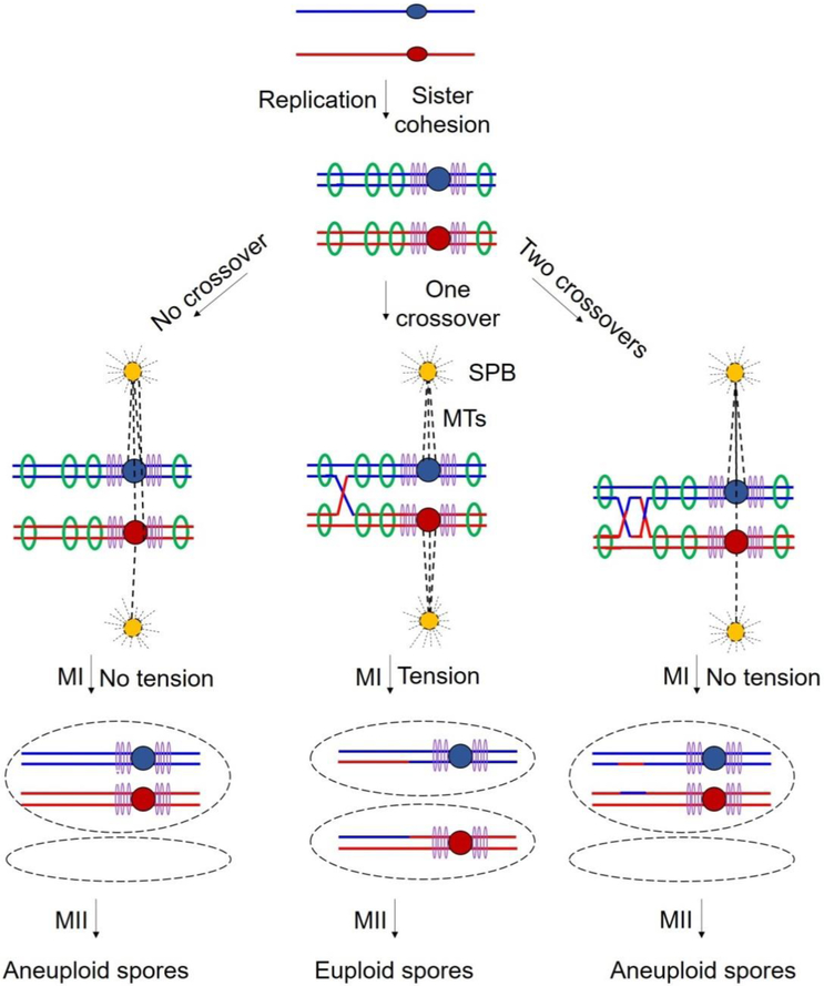
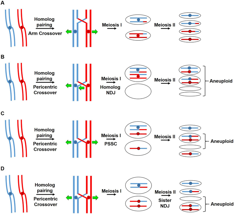
During meiosis, homologous chromosomes of a diploid cell are replicated and, without a second replication, are segregated during two nuclear divisions to produce four haploid cells (including discarded polar bodies in females of many species). Proper segregation of chromosomes at the first division requires in most species that homologous chromosomes be physically connected. Tension generated by connected chromosomes moving to opposite sides of the cell signals proper segregation. In the absence of the required connections, called crossovers, chromosomes often segregate randomly and produce aneuploid gametes and, thus, dead or disabled progeny. To be effective, crossovers must be properly distributed along chromosomes. Crossovers within or too near the centromere interfere with proper segregation; crossovers too near each other can ablate the required tension; and crossovers too concentrated in only one or a few regions would not re-assort most genetic characters important for evolution. Here, we discuss current knowledge of how the optimal distribution of crossovers is achieved in the fission yeast Schizosaccharomyces pombe, with reference to other well-studied species for comparison and illustration of the diversity of biology.

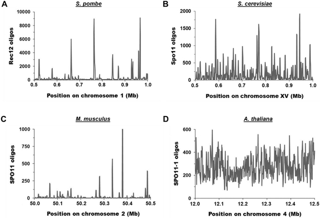
Keywords: Crossover interference; Crossover invariance; DNA break hotspots; DNA break interference; Homologous recombination; Linear element proteins; Meiosis; Pericentric repression; Schizosaccharomyces pombe.


Conflict of interest statement

Conflict of Interest

The authors declare no conflict of interest.

Figures

Figure 1.
Figure 1.. Pathway of meiotic recombination in S. pombe (modified from [93]).
Thick lines indicate the ds DNA of a chromatid, red for the chromosome from one parent and blue from the other parent; black dots indicate the centromeres. In ovals, thin lines indicate a single strand of DNA; dotted red lines indicate newly synthesized DNA. Accompanying replication, sister chromatid cohesins, containing the meiosis-specific subunits Rec8 and Rec11, are loaded onto chromosomes. In chromosomal arms casein kinase I (CK1; Hhp1 and Hhp2) phosphorylates Rec8, for its proteolysis and sister chromatid segregation, and Rec11, for its recruiting Rec10. Rec25, Rec27, and Mug20 direct Rec10 at high frequency to DSB hotspots. Rec10 binds Rec15, which with other indicated proteins activates Rec12 (Spo11 homolog) to make DSBs. The MRN complex and Ctp1 clip off Rec12 covalently bound to 5’ DNA ends and further resect the 5’ ends to produce long 3’-ended ss DNA tails. Rad51 and Dmc1 DNA strand-exchange proteins bind the tails and, with the additional proteins listed, form a displacement- (D-) loop with intact ds DNA. The D-loop (not shown) is cleaved to form a Holliday junction (HJ), which is resolved by the Mus81-Eme1 complex into crossover (shown) or non-crossover (gene conversion; not shown) products. HJ resolution is aided in an unknown way by Nse5 and Nse6, subunits of the Smc5-Smc6 complex [94]. Additional gene products required for meiotic recombination, but whose point of action remains unknown, include the following: mug1 (jnm1), mug5, pds5, rad54, rdh54, and rlp1; hop1 and mek1 [95], nse1 [96], pli1 [97], rec13, rec18, and rec21 [98], and rqh1 [99]. See [93] for references for genes not otherwise referenced here.
Figure 2.
Figure 2.. Crossovers are essential for proper segregation.
Each line indicates the ds DNA of a chromatid, red for the chromosome from one parent and blue from the other parent; central dots indicate the centromeres. After replication, sister chromatids of each parent are held together by cohesin complexes deposited at points across the chromosomal arms (green rings) and especially densely at the pericentric regions (purple rings). At MI, paired centromeres of each homolog attach to microtubules (MTs; dashed lines) originating from spindle pole bodies (SPBs) at opposite poles of the cell. Middle panels: Proper reductional division at meiosis I (MI) results from tension generated by centromeres being pulled to opposite poles by MTs and dependent on both cohesion between sister chromatids and crossovers between homologs. Left panels: Without crossovers, no tension is generated when MTs begin to pull the homologs to opposite poles. Right panels: Crossovers too close together may lack intervening cohesion and thus no tension being generated.
Figure 3.
Figure 3.. Distribution of Spo11 oligos (DSBs) across representative 0.5 Mb chromosomal arm regions in four species.
The number of Spo11 oligos per 1 kb bin is plotted for each representative genomic region. (A) S. pombe [33]. The mean and median values are 339.9 and 81.5, respectively. (B) S. cerevisiae [32]. The mean and median values are 177.1 and 76.5, respectively. (C) M. musculus [34]. The mean and median values are 19.5 and 2.1, respectively. (D) A. thaliana [35]. The mean and median values are 272.6 and 257.0, respectively.
Figure 4.
Figure 4.. Positioning of crossovers on meiotic chromosomes dictates proper segregation and correct ploidy in gametes.
Each line indicates the ds DNA of a chromatid, red for the chromosome from one parent and blue from the other parent; central dots indicate the centromeres. Crossovers in the chromosomal arms (depicted as red-blue junctions) are needed for accurate chromosomal segregation during meiosis (A) (see Figure 2), but crossovers too close to the centromere are deleterious. Pericentric crossovers can lead to homolog non-disjunction (NDJ) at MI (B), precocious separation of sister chromatids (PSSC) when cohesion is lost between the sister chromatids at MI (C), or sister NDJ at MII (D). These missegregation events lead to aneuploid spores, either nullisomic (lacking a chromosome) or disomic (having an extra chromosome).
Figure 5.
Figure 5.. Composition of the cohesin complexes differentially controls distribution of DSB formation and recombination.
Each solid line indicates the ds DNA of a chromatid. In euchromatic DNA present largely in chromosomal arms, the meiosis-specific Rec8 and Rec11 subunits are part of the cohesin complex (with Smc1-Smc3). Rec11 activates Spo11- (Rec12)-dependent DSB formation by recruiting the LinE protein Rec10 (Figure 1). Other LinE proteins Rec25, Rec27 and Mug20 are recruited with Rec10 at DSB hotspots. Rec10 activates the Spo11 complex to induce DSB formation. In contrast, in heterochromatic DNA, especially at pericentric regions, the cohesin complex contains Rec8-Psc3, which does not bind Rec10 or activate DSB formation. Loading of Rec8-Psc3 cohesin complex is mediated by the HP1 homolog Swi6, which specifically binds to H3K9 methylated histones present in heterochromatin. Note that Swi6 has both a negative role (recruiting Psc3) and a potentially positive role (recruiting Rec8 via Psc3 recruitment); thus, swi6 mutants are not derepressed for pericentric recombination. The scheme shown is for S. pombe and is likely conserved even in mammals (see text).
Figure 6.
Figure 6.. A molecular model for crossover interference – the clustering model.
(A) Live cell image of LinE foci at the "horsetail" stage of S. pombe meiosis. Multiple LinEs (represented by Rec25-GFP) concentrate and form linear or dot-like foci. DNA stained by Hoechst 33342 is red, and Rec25-GFP is green; overlap is yellow. (B-C) Model of crossover interference based on hotspot clustering and DSB interference. Each horizontal line is one sister chromatid (ds DNA molecule). Ovals indicate clusters, within which one DSB (yellow lightning bolt) occurs. (B) DSB hotspots are bound by LinEs (green dots) and form clusters of nearby hotspots on sister chromatids. Only one DSB and hence one crossover are formed per cluster. Independent cluster and DSB formation on separate homologs allows close double crossovers and thus low but positive crossover interference. (C) Clusters encompass all four chromatids in species with strong crossover interference. (D) Clusters encompass only two sister chromatids (one homolog) in species with weak crossover interference. Figure modified from [82].

References

    1. Powsner ER, Berman L, Number of chromosomes in the human cell, Nature, 188 (1960) 1045–1046. - PubMed
    1. Sinha B, Srivastava D, Jha J, Occurrence of various cytotypes of ophioglossum reticulatum L. in a population from N. E. India, Caryologia, 32 (1979) 135–146.
    1. Goday C, Pimpinelli S, Chromosome organization and heterochromatin elimination in parascaris, Science, 224 (1984) 411–413. - PubMed
    1. Crosland MW, Crazier RH, Myrmecia pilosula, an ant with only one pair of chromosomes, Science, 231 (1986) 1278. - PubMed
    1. Davis L, Rozalén AE, Moreno S, Smith GR, Martin-Castellanos C, Rec25 and Rec27, novel components of meiotic linear elements, link cohesin to DNA breakage and recombination in fission yeast, Current Biology, 18 (2008) 849–854. - PMC - PubMed

Publication types

LinkOut - more resources